Conjunctival Nevus: A Benign Growth on the Eye's Surface
What Is Conjunctival Nevus?
Peering into the world with bright, expressive eyes is a joy that can captivate hearts and ignite curiosity. However, when a benign growth like a conjunctival nevus takes residence on the surface of the eye, it introduces a unique twist to the ocular landscape. Conjunctival nevi, like tiny brushstrokes of pigmented or non-pigmented artistry, add a touch of intrigue to the conjunctiva.
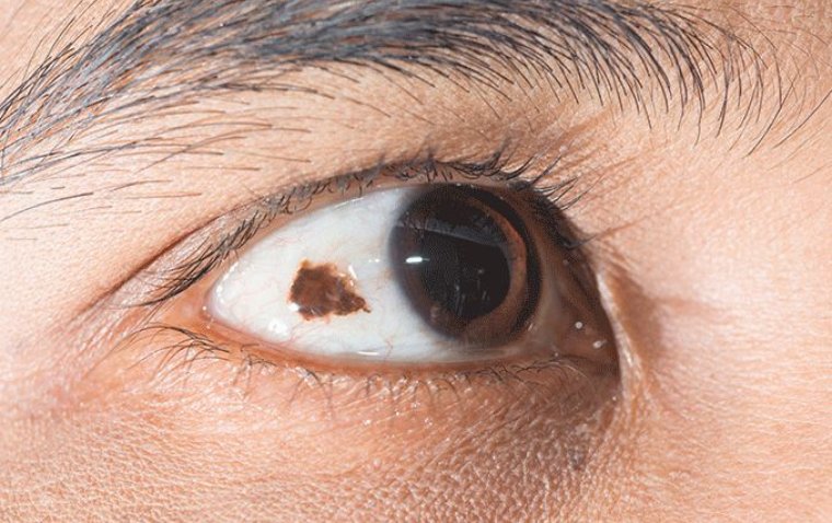
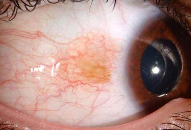
Conjunctival nevus refers to a common benign growth that develops on the conjunctiva, which is the clear, thin membrane that covers the sclera (white part) of the eye and lines the inner surface of the eyelids. This condition is characterized by the presence of pigmented or non-pigmented elevated lesions on the surface of the conjunctiva.
Conjunctival nevi are typically harmless and do not pose a significant threat to vision or overall eye health. However, regular monitoring and evaluation by an ophthalmologist are essential to ensure early detection of any changes and to distinguish them from potentially malignant conjunctival melanoma. Let's dive deeper into understanding this fascinating ocular phenomenon.
Causes and Risk Factors of Conjunctival Nevus
The exact cause of conjunctival nevus formation is not fully understood. However, several factors may contribute to their development. Here, we explore the underlying causes and potential risk factors associated with conjunctival nevi.
Underlying Causes of Conjunctival Nevus
Melanocytic Proliferation
Conjunctival nevi originate from the proliferation of melanocytes, which are pigment-producing cells present in the conjunctiva. The precise mechanism triggering the abnormal growth of these cells is still under investigation.
Genetic Mutations
Genetic alterations in certain genes involved in the regulation of cell growth and differentiation have been implicated in the development of conjunctival nevi. However, the specific genes involved and the mechanisms through which they contribute to nevus formation are yet to be fully elucidated.
Risk Factors for Conjunctival Nevus
While the development of conjunctival nevi can occur in anyone, certain risk factors may increase the likelihood of their occurrence. These factors include:
● Sun exposure: Prolonged and unprotected exposure to ultraviolet (UV) radiation from sunlight is considered a significant risk factor for conjunctival nevus formation. UV radiation can induce genetic changes in the conjunctival cells, leading to the development of nevi.
● Fair skin and light-colored eyes: Individuals with fair skin and light-colored eyes are more susceptible to the harmful effects of UV radiation. As a result, they may have an increased risk of developing conjunctival nevi.
● Genetic predisposition: There is evidence to suggest that a genetic predisposition may play a role in the development of conjunctival nevi. Individuals with a family history of nevi or other pigmented lesions may have a higher risk of developing conjunctival nevi themselves.
● Age: Conjunctival nevi typically appear during childhood or adolescence, although they can develop at any age. The likelihood of developing nevi may decrease with advancing age.
How to Diagnose Conjunctival Nevus
Accurate diagnosis of conjunctival nevus is essential to differentiate it from other ocular conditions and determine appropriate management strategies. The diagnostic process typically involves a comprehensive evaluation that incorporates various examination techniques. Here, we outline the key steps involved in diagnosing conjunctival nevi.
Visual Examination
● External Eye Examination: A thorough examination of the external eye and eyelids is conducted to assess any visible signs or abnormalities on the conjunctiva. The size, shape, color, and location of the nevus are carefully observed.
● Slit Lamp Examination: A slit lamp biomicroscope is used to examine the nevus in more detail. This instrument provides a magnified view of the conjunctiva and enables the ophthalmologist to assess the surface characteristics of the nevus, such as its texture, pigmentation, and vascularity.
Imaging Techniques
● Anterior Segment Photography: High-resolution photography using specialized equipment allows for detailed documentation of the conjunctival nevus. Serial photographs taken over time can aid in monitoring any changes or growth of the lesion.
● Confocal Microscopy: This non-invasive imaging technique provides real-time, high-resolution images of the conjunctiva at a cellular level. It can help in assessing the cellular structure of the nevus and distinguishing it from other ocular conditions.
● Optical Coherence Tomography (OCT): OCT utilizes light waves to capture cross-sectional images of the conjunctiva. It can provide valuable information about the thickness and depth of the nevus, as well as its relationship with the underlying layers of the eye.
Importance of Differentiation
It is crucial to differentiate conjunctival nevi from other ocular conditions, particularly potentially malignant conjunctival melanoma. The following factors aid in distinguishing conjunctival nevi from other lesions:
● Clinical Appearance: Conjunctival nevi typically have well-defined borders, uniform pigmentation, and smooth surfaces. Other conditions may exhibit irregularities, asymmetry, or rapid growth.
● Imaging Findings: Advanced imaging techniques like confocal microscopy and OCT can provide additional insights into the cellular characteristics and depth of the lesion, helping to differentiate nevi from other conditions.
● Patient History: A thorough evaluation of the patient's medical history, family history, and any relevant symptoms can provide valuable information in the diagnostic process.
If there is uncertainty in the diagnosis or suspicion of malignancy, a biopsy may be performed to obtain a sample for histopathological examination.
Proper diagnosis of conjunctival nevus allows for appropriate monitoring and management, ensuring the preservation of ocular health and providing peace of mind for patients. Regular follow-up examinations are recommended to monitor any changes in the nevus over time.
Treatment and Management of Conjunctival Nevus
The management of conjunctival nevus primarily depends on the characteristics of the lesion, its impact on vision, and the potential risk of malignancy. In many cases, conjunctival nevi are benign and require regular monitoring and observation. However, in certain situations, intervention may be necessary. Here, we discuss the various treatment options available for conjunctival nevus.
Regular Monitoring and Observation
Periodic Eye Examinations: Regular follow-up examinations with an ophthalmologist are crucial to monitor any changes in the size, shape, or appearance of the nevus over time. Typically, follow-up intervals can range from several months to years, depending on the individual case.
Photographic Documentation: Serial anterior segment photography allows for accurate documentation and comparison of the nevus over time, aiding in the detection of any subtle changes.
Treatment Options
Surgical Excision
Surgical removal of the conjunctival nevus may be considered in cases where the lesion causes significant symptoms, affects vision, or shows signs of potential malignancy. The excision is typically performed under local anesthesia, and the extent of tissue removal depends on the size and location of the nevus. The excised tissue is sent for histopathological examination to confirm the diagnosis and rule out any malignant transformation.
Cryotherapy
Cryotherapy involves freezing the nevus using extreme cold temperatures. It is often utilized for small and superficial nevi. Cryotherapy causes controlled cell destruction and subsequent healing of the treated area. This technique may be combined with surgical excision for certain cases.
Other Techniques
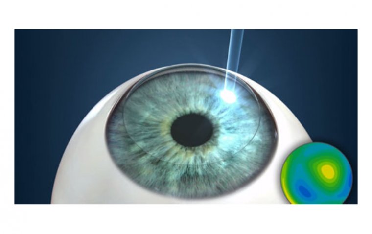
Alternative treatment options, such as laser therapy or radiotherapy, have been explored in select cases. However, their use is relatively limited and requires careful consideration on a case-by-case basis.
The choice of treatment depends on various factors, including the size, location, and characteristics of the nevus, as well as the patient's overall health and preferences. The goal of treatment is to preserve vision, alleviate symptoms, and ensure the safety of the individual.
Prognosis and Follow-up
Conjunctival nevi, in general, have a favorable prognosis and tend to remain stable or exhibit slow growth over time. These benign lesions rarely cause significant visual impairment or discomfort. However, regular follow-up visits with an eye care professional are crucial to monitor the nevus for any changes and ensure early detection of potential complications. Here, we discuss the typical prognosis of conjunctival nevi and the importance of ongoing surveillance.
Prognosis of Conjunctival Nevus
Stability and Slow Growth: Conjunctival nevi often exhibit a stable course and demonstrate slow growth, if any, over an extended period. They are considered benign and do not pose an immediate threat to vision or overall ocular health.
Low Risk of Malignant Transformation: While conjunctival nevi have a generally low risk of transforming into malignant melanoma, it is essential to recognize that this transformation is possible, albeit rare. Regular monitoring helps detect any signs of suspicious changes that may indicate malignant transformation.
Importance of Regular Follow-up Visits
Detection of Changes
Regular follow-up visits with an eye care professional, such as an ophthalmologist or optometrist, allow for close monitoring of the conjunctival nevus. Any changes in size, shape, pigmentation, or symptoms can be promptly identified and evaluated.
Early Detection of Malignant Transformation
Although rare, there is a potential risk of conjunctival nevi transforming into malignant melanoma. Regular follow-up visits help in the early detection of any signs suggestive of malignant transformation, such as rapid growth, irregular borders, asymmetry, or changes in color.
Optimal Treatment Planning
Continuous monitoring enables the eye care professional to assess the need for further interventions, such as surgical excision, cryotherapy, or referral to a specialist if there is suspicion of malignancy. Early intervention improves the chances of successful treatment and favorable outcomes.
Patient Education and Support for Conjunctival Nevus
Patient education plays a vital role in empowering individuals with conjunctival nevi to understand their condition, make informed decisions, and take proactive steps for their ocular health. Comprehensive patient education should cover various aspects of conjunctival nevi, including their benign nature, common characteristics, and measures to promote ocular health. Here, we discuss the importance of providing patient education and support for individuals with conjunctival nevi.
Understanding Benign Nature
Patients should be informed that conjunctival nevi are typically benign growths on the surface of the eye. Emphasize that most nevi remain stable or exhibit slow growth over time and do not pose an immediate threat to vision or overall ocular health.
Common Characteristics
Educate patients about the typical features of conjunctival nevi, such as pigmented or non-pigmented elevated or flat lesions on the conjunctiva. Explain that nevi can vary in size, color, and appearance, and may or may not cause symptoms.
Sun Protection
Highlight the importance of sun protection for overall ocular health. Encourage patients to wear sunglasses that provide UV protection and to use hats or visors to shield their eyes from direct sunlight, as excessive sun exposure is considered a potential risk factor for conjunctival nevi.
Regular Eye Examinations
Stress the significance of regular eye examinations with an eye care professional. Explain that routine check-ups enable the monitoring of the nevus for any changes and early detection of potential complications. Encourage patients to adhere to their scheduled follow-up visits.
Supporting Measures for Ocular Health
● Self-Examination: Teach patients how to perform self-examinations of their conjunctival nevi. Encourage them to regularly observe and note any changes in size, color, or symptoms. Advise them to report any concerning changes to their eye care professional.
● Awareness of Risk Factors: Provide information about potential risk factors associated with conjunctival nevi, such as sun exposure or genetic predisposition. Patients should be aware of these factors and take appropriate preventive measures.
● Addressing Concerns: Create an environment where patients feel comfortable discussing their concerns and seeking clarification. Address any misconceptions or anxieties they may have about their condition. Clear communication helps alleviate fears and promotes better patient engagement.
● Psychosocial Support: Recognize the emotional impact conjunctival nevi may have on patients. Offer psychosocial support, including reassurance, empathy, and resources for additional counseling or support groups, if needed.
Conclusion
In conclusion, conjunctival nevi, these benign adornments on the eye's surface, are a fascinating aspect of ocular health. With comprehensive patient education and support, individuals with conjunctival nevi can gain a deeper understanding of their condition, embrace its benign nature, and actively participate in maintaining their ocular well-being. By emphasizing sun protection, regular eye examinations, and self-awareness, we can ensure the long-term health of the eye and promptly address any changes or concerns. As we continue to unravel the complexities of conjunctival nevi, let us strive to provide the care and support needed to enhance the lives of those with these captivating ocular companions.
FAQ
Yes, conjunctival nevi can grow, change in size, shape, or color, requiring regular monitoring.
(1).jpg)
