A Step-by-Step Guide to Visual Field Testing for Residents
What Is Visual Field Testing?
Visual field testing is a vital diagnostic procedure used to evaluate the extent and quality of a person's peripheral vision. It helps detect and monitor various ocular and neurological conditions, such as glaucoma, retinal disorders, and brain tumors. This article aims to provide a comprehensive guide on how to conduct visual field testing, outlining the equipment, techniques, and interpretation of results.
Different Testing Techniques
1. The Goldmann Perimetry: This manual technique employs a bowl-like instrument called a perimeter. It assesses the visual field by measuring the smallest target size a person can detect in different locations.
2. Automated Perimetry: Utilizing a computerized device, this technique presents stimuli in a predetermined pattern, assessing a person's response to light.
3. Kinetic Perimetry: Involves moving an object from the periphery to the center of vision, determining the point at which the person perceives it.
4. Static Perimetry: Displays a grid of light stimuli on a screen, and the person responds when they see the light appear.
Preparing for Visual Field Testing
Performing Visual Field Testing
Performing visual field testing involves several crucial steps to obtain accurate and reliable results.
1. Explain the test procedure to the patient, ensuring they understand the importance of concentration and fixation.
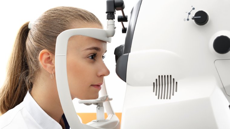
2. Correctly position the patient by aligning their eyes with the visual field instrument.
3. Instruct the patient to fixate on a central target during the entire test.
4. For automated perimetry, ensure proper alignment and patient focus.
5. Establish the appropriate test parameters based on the patient's condition, such as stimulus size, duration, and intensity.
6. Conduct a practice test or threshold determination to acquaint the patient with the process.
Interpreting Visual Field Test Results
Interpreting visual field test results requires a comprehensive analysis of the data obtained. The visual field indices provided by the testing instrument, such as mean deviation (MD), pattern standard deviation (PSD), and visual field index (VFI), offer valuable information about the patient's visual field.
Comparing the results to age-matched norms or previous tests helps identify any deviations or changes over time. It is crucial to pay attention to visual field defects, such as scotomas (blind spots), generalized depression, or localized abnormalities. Analyzing the pattern and symmetry of visual field defects aids in differentiating between ocular and neurological causes.
To arrive at an accurate interpretation, it is essential to correlate the visual field findings with the patient's clinical history, symptoms, and other diagnostic tests. This comprehensive approach allows healthcare professionals to determine the underlying condition and guide appropriate treatment and management strategies.
Additional Considerations While Interpreting Visual Field Test Results
When interpreting visual field testing results, it is important to take into account several additional considerations. First, understanding that practice and experience play a significant role in accurately interpreting the results is crucial. Familiarity with normal variations and patterns of visual field defects helps distinguish between physiological variations and pathological conditions.
It is also essential to consider factors that can influence the results, such as patient fatigue, learning effects, and cooperation. Patient compliance and concentration during the test can greatly impact the reliability of the results. Additionally, staying updated with the latest advancements and technologies in visual field testing is important to enhance interpretation skills.
Regularly attending educational programs and staying informed about new research findings will ensure healthcare professionals can confidently interpret visual field test results and provide optimal care for their patients.
Conclusion
Visual field testing is an essential diagnostic tool for evaluating and monitoring various ocular and neurological conditions. By following the guidelines outlined in this comprehensive guide, healthcare professionals can conduct accurate visual field tests, interpret the results effectively, and contribute to the early detection and management of visual disorders. Remember, consistent practice and ongoing education are key to mastering this valuable clinical skill.
(1).jpg)
