(1).jpg)

Understanding Optic Atrophy: Impaired Vision and Beyond
What Is Optic Atrophy?
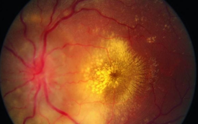
Optic atrophy refers to an end-stage destruction of the retinal ganglion cell axons that comprise the optic nerve causing impairment to the anterior visual pathway (the path from the retina to the lateral geniculate body). On fundoscopy, it appears as pale optic disc. Optic atrophy and optic neuropathy are used interchangeably as well.
Optic atrophy cannot be regarded as a disease in itself, it is a clinical manifestation of some underlying disease.
Causes of Optic Atrophy
●Anterior ischemic optic neuropathy caused by stroke of the optic nerve
●Glaucoma
●Optic nerve compression by a tumor
●Optic neuritis due to multiple sclerosis
●Leber's hereditary optic neuropathy
●Congenital malformation of the optic nerve
Signs and Symptoms of Optic Atrophy
●Loss of vision
●Blurring or hazy vision
●Impaired peripheral (side) vision
●Inconveniencies with color vision
●A decrease in sharpness of the vision
●Diminished sharpness in vision
Visual Field Defects
●Visual field patterns consist of papillomacular defect
●Arcuate defect or temporal wedge defect (nasal fibers) for prechiasmal lesions
●Bitemporal (superior) field defects due to chiasmal lesions
●Hemianopsia in post-chiasmal lesions.
How to Diagnose Optic Atrophy
History
History plays an indispensable role in precise diagnosis of medical conditions. Ophthalmologists need to know the history of patients presenting complaints, medications, other diseases, ocular trauma as well as previous visits to hospital for similar complaints.
Physical Examination
Next step is complete eye exam of the patient by the ophthalmologist, paying close attention to: -
●Visual Field tests
●Color Vision Assessment
●Contrast Vision Assessment
●Intra Ocular Pressure in Eyes
●RAPD
●Fundoscopy
Clinical Diagnosis
Hallmark of optic atrophy for clinical diagnosis is pale optic disc on fundoscopy. The root cause for the optic atrophy may be challenging to determine. Numerous tests are available to achieve this purpose: -
●Electrophysiology (ERG, mERG) to exclude the probability of retinal diseases.
●OCT to measure the thinning of the peripapillary retinal nerve fiber layer.
●Visual Field Testing to help localize the site of the lesion.
●Neuro-imaging (MRI, CT) to judge for compressing factors like tumors or bone growth other than that fractures and multiple sclerosis can also be identified.
●Carotid Doppler ultrasound.
●Laboratory screening tests are done for B12 level, folate level, VDRL, ACE and Antiphospholipid antibodies after enough evidence of these on history.
Is There a Cure for Optic Atrophy?
There is no pharmacological treatment available for optic atrophy.
The intention is to protect residual function of optic nerve by early detection before the irrevocable optic atrophy is noted. Research studies show that the optic nerve has several reserves (axons) before significant vision loss takes place. If this reserve is depleted slight changes in nerve fibers leads to noteworthy diminution in vision. For this purpose, identification and treatment of underlying causes of optic atrophy like multiple sclerosis and glaucoma are crucial.
Restoring the dead axons is currently not a possibility consequently there is no available treatment for optic atrophy, however, research is being done on stem cell treatment for optic atrophy which might prove to be key in future treatment.