(1).jpg)
What Does It Mean to Have Cotton Wool Spots on the Retina?
What Are Cotton Wool Spots?
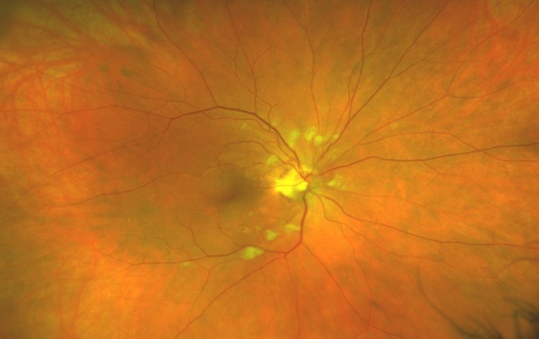
Cotton Wool Spots (CWS) are small, white or grayish lesions on the retina—the layer of cells at the back of the eye responsible for converting light into neural signals. These spots signify local ischemia, where blood flow to the retinal nerve fibers is reduced or obstructed, leading to their swelling and eventual necrosis. They are commonly identified during an eye examination using an ophthalmoscope and often manifest as fluffy white patches on the retina.
CWS are not a disease in themselves but rather a symptom that indicates a range of potential underlying systemic health issues. They are particularly associated with conditions that affect the vascular system, such as diabetes and hypertension. For instance, the presence of a single cotton wool spot can be one of the first signs of diabetic or hypertensive retinopathy, and in some studies, patients with CWS have shown a significantly higher prevalence of elevated diastolic blood pressure.
While CWS often do not directly affect vision and may resolve without specific treatment, they should not be disregarded, as they can be a harbinger of more serious medical conditions that might have systemic and ocular consequences if left untreated. The potential consequences of untreated CWS can include the progression to more severe retinopathy, an increased risk of stroke, and other cardiovascular complications due to the vascular diseases they may signify. Therefore, the detection of CWS calls for a comprehensive medical evaluation to determine and manage the underlying cause effectively.
What is the Difference between Exudates and Cotton Wool Spots?
Cotton wool spots (CWS), sometimes referred to as "soft exudates," are small, slightly elevated, yellow-white or gray-white lesions on the superficial retina. They have a cloud-like, linear, or serpentine appearance with fimbriated borders and are typically perpendicular to the nerve fibers in the retina, with a fluffy border and a whitish color.
In contrast, exudates, particularly hard exudates found in conditions like diabetic retinopathy, are made of lipid and proteinaceous material such as fibrinogen and albumin. They result from leakage from an impaired blood-retinal barrier and are primarily deposited in the outer plexiform layer of the retina. Hard exudates appear yellowish and granular, and when they accumulate in the macular region, they can cause significant visual loss.
Causes and Risk Factors
Cotton wool spots (CWS) are retinal lesions that typically represent local ischemia, a condition where there is an inadequate blood supply to the retinal nerve fibers due to obstruction or narrowing of the retinal blood vessels. This ischemia leads to the swelling and subsequent necrosis of nerve fibers, appearing as fluffy white patches on the retina during an eye examination. CWS are not diseases themselves but are indicative of underlying conditions, most notably diabetes mellitus and systemic hypertension.
The development of CWS can be tied to several causes and risk factors:
1. Vascular Issues: Conditions that affect blood vessels, such as hypertension, can cause damage to the retinal blood vessels over time. Long-term and uncontrolled high blood pressure can lead to hypertensive retinopathy, where the damaged retinal vessels can result in CWS as part of the disease spectrum.
2. Diabetes Mellitus: Diabetes is a significant risk factor for CWS because of the vascular complications associated with the disease. Diabetic retinopathy often involves the formation of CWS due to the damage diabetes causes to the blood vessels of the retina.
3. Other Systemic Diseases: CWS can also be associated with a range of other systemic diseases such as connective tissue diseases, neoplastic conditions, and infectious diseases. In these cases, the CWS are a manifestation of the broader impact these illnesses have on the vascular system and particularly on the small vessels that nourish the retina.
4. Occlusion of Retinal Arterioles: A single CWS may result from an acute focal occlusion of a single retinal arteriole. This event can be related to the systemic conditions mentioned above or to localized retinal vascular issues.
Given these risk factors, it is evident that CWS are a marker for systemic health and should prompt further investigation into the patient's overall vascular health. Managing the underlying conditions, such as controlling blood sugar in diabetes or managing blood pressure in hypertension, is critical to preventing the development of CWS and the potential complications they signify.
.jpg)
Symptoms and Diagnostic Process
Cotton wool spots (CWS) often present asymptomatically, meaning patients may not notice any signs. However, when symptoms do occur, they can include varying degrees of blurry vision and, more rarely, visual field defects such as scotomas (areas of partial alteration in the field of vision), arcuate defects, or transient vision loss known as amaurosis fugax.
The diagnostic process for CWS begins with a thorough history and eye exam, which includes a dilated retinal evaluation to inspect the retinal nerve fiber layer where CWS manifest. In addition to the physical exam, further workup might include blood tests to explore other potential causes. These tests can range from a complete blood count, a basic metabolic panel, to more specific tests for digestive enzymes, and several blood pressure measurements. Spectral domain optical coherence tomography (SD-OCT) is another advanced diagnostic tool that can be used to monitor the progression of CWS and provide detailed images of the retinal layers.
In the clinical setting, CWS are characterized by localized, white-yellowish, fluffy areas of nerve fiber layer edema caused by focal ischemia that interrupts axoplasmic flow, leading to the accumulation of axoplasmic content within the retina. This appearance is distinct from other retinal conditions, allowing clinicians to identify CWS as part of a broader diagnostic workup when patients present with retinal issues or systemic symptoms that might be related to vascular diseases.
Management of Cotton Wool Spots
The management of Cotton Wool Spots (CWS) primarily involves addressing the underlying etiology, as CWS themselves usually resolve within 6–12 weeks. In conditions such as diabetic retinopathy, however, they may persist longer. The initial workup to identify the cause may include checking vitals, such as blood pressure and heart rate, and conducting metabolic studies, including glycated hemoglobin (HbA1c), complete blood count (CBC), comprehensive metabolic panel (CMP), and HIV tests. Further directed workups may involve inflammatory markers like ESR and CRP, cardiovascular assessments with EKG and echocardiogram, carotid ultrasound, hypercoagulable labs, and homocysteine levels.
Regular eye check-ups are vital for managing CWS, especially for individuals with known risk factors like diabetes and hypertension. These check-ups can help monitor the resolution of CWS and detect any potential recurrence or progression of the underlying conditions that caused them.
Summary
In summary, Cotton Wool Spots (CWS) are retinal changes indicative of localized ischemia, often associated with systemic conditions like diabetes and hypertension. While they can be asymptomatic, CWS might lead to blurry vision and should be evaluated for underlying causes. Management focuses on treating these conditions and includes regular monitoring of the spots through eye examinations. The disappearance of CWS can typically be expected, but persistent spots, especially in diabetic patients, require a comprehensive medical approach. Regular eye check-ups are crucial for early detection and management of any retinal changes, including CWS.
FAQ