(1).jpg)
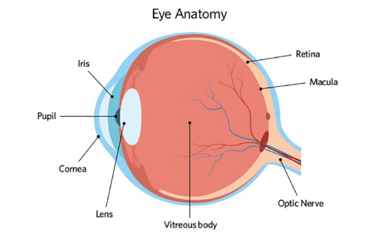
The Eye: An In-Depth Look at Its Incredible Anatomy
The human eye is one of the most complex and important sensory organs in the body. It allows us to see and interpret the world around us. The anatomy of the eye is made up of several different parts that work together to help us see. In this article, we will explore the main parts of the human eye.
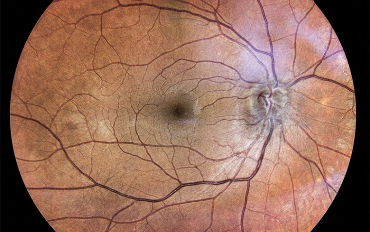
● Retina
The retina is a thin layer of tissue that lines the back of the eye. It is responsible for receiving and processing light that enters the eye. The retina contains two types of cells that are sensitive to light: rods and cones. Rods are responsible for detecting light levels, while cones are responsible for detecting color. The retina is connected to the brain via the optic nerve, which transmits visual information to the brain for processing.
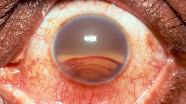
● Iris
The iris is the colored part of the eye that surrounds the pupil. It is made up of muscles that control the size of the pupil, which regulates the amount of light that enters the eye. The iris also plays a role in determining the color of the eyes. The iris can change the size of the pupil in response to changes in light levels, allowing the eye to adjust to different lighting conditions.
● Macula
The macula is a small, oval-shaped area near the center of the retina. It is responsible for providing sharp, detailed vision. The macula contains a high concentration of cones, which are responsible for detecting color and fine details. The macula is essential for activities that require fine visual discrimination, such as reading and driving.
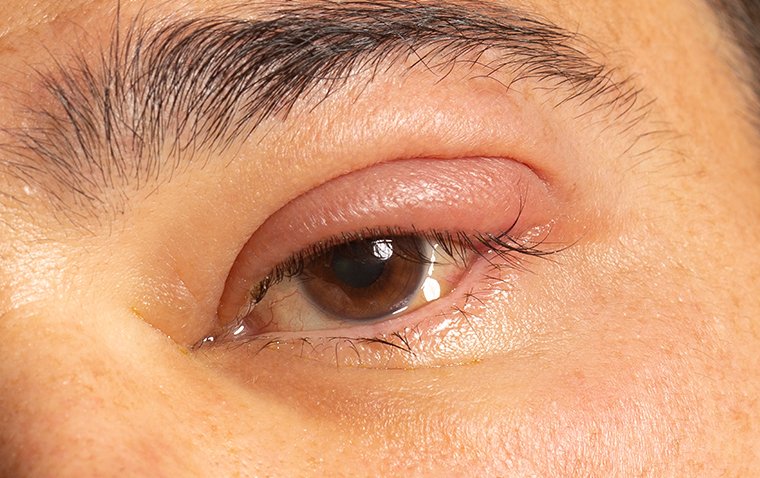
● Eyelids
The eyelids are two thin folds of skin that cover and protect the eye. They help to keep the eye moist by distributing tears across the surface of the eye. The eyelids also play a role in regulating the amount of light that enters the eye. The eyelids can close rapidly to protect the eye from foreign objects or bright light.
● Lacrimal Gland
The lacrimal gland is a gland located in the upper outer corner of each eye socket, behind the upper eyelid. It is responsible for producing tears, which are important for lubricating and protecting the surface of the eye.
Tears are composed of a mixture of water, mucus, and other substances, such as antibodies and enzymes, that help to keep the surface of the eye moist and free from debris. The lacrimal gland produces the watery component of tears, while other glands in the eyelids produce the mucus and oil components.
● Cornea
The cornea is the clear, dome-shaped outer layer of the eye. It is responsible for refracting, or bending, light that enters the eye. The cornea is also responsible for protecting the eye from foreign objects and injury. The cornea is the first structure that light encounters when it enters the eye, and it is responsible for the majority of the eye's refractive power.
● Ciliary Body
The ciliary body is a structure in the eye that is located between the iris (the colored part of the eye) and the choroid (the layer of blood vessels that nourishes the retina). It is a ring-shaped structure that is responsible for several important functions in the eye.
● Aqueous Humor
Aqueous humor is a clear, watery fluid that fills the space between the cornea (the clear outer covering of the eye) and the lens of the eye. It is produced by a structure called the ciliary body. There are two different types of aqueous humor: the anterior chamber and the posterior chamber. The anterior chamber is located between the cornea and iris, while the posterior chamber is located between the iris and lens.
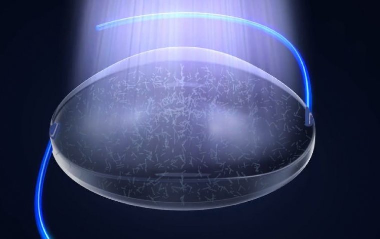
● Vitreous Humor
The vitreous humor is a clear, gel-like substance that fills the space between the lens of the eye and the retina, the light-sensitive tissue at the back of the eye. It makes up about two-thirds of the volume of the eye and is responsible for maintaining the shape of the eyeball.
The vitreous humor is composed of 99% water and a network of collagen fibers and other proteins that give it its gel-like consistency. It also contains various nutrients and molecules that support the health and function of the retina.
● Lens
The lens is a transparent, flexible structure that sits behind the iris. It is responsible for focusing light onto the retina. The lens changes shape to adjust the focus of the eye, allowing us to see objects at different distances. The ability of the lens to change shape is called accommodation. As we age, the lens becomes less flexible, and it becomes more difficult to focus on near objects, leading to a condition known as presbyopia.
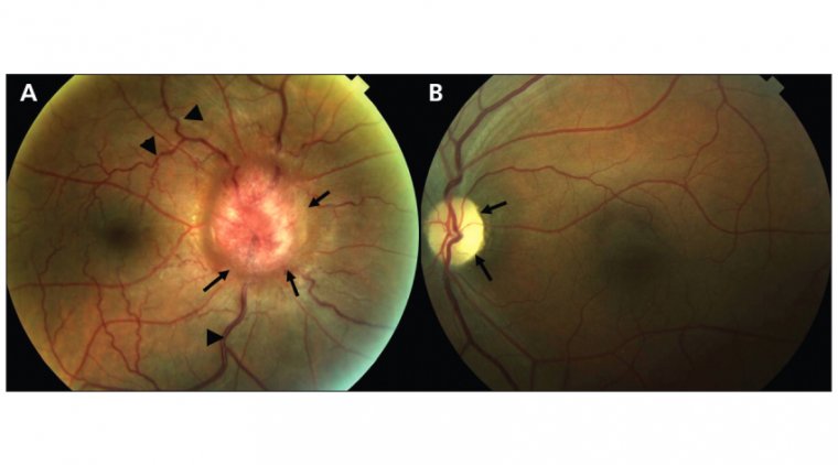
● Optic Nerve
The optic nerve is a bundle of nerve fibers that carries visual information from the retina to the brain. The brain processes this information to create the images that we see. The optic nerve is made up of approximately one million nerve fibers, and damage to the optic nerve can lead to vision loss or blindness.
● Conjunctiva
The conjunctiva is a thin, transparent layer of tissue that covers the front of the eye and lines the inside of the eyelids. It helps to protect the eye from infection and lubricates the eye by producing tears. The conjunctiva is highly vascular and can become inflamed, leading to a condition known as conjunctivitis.
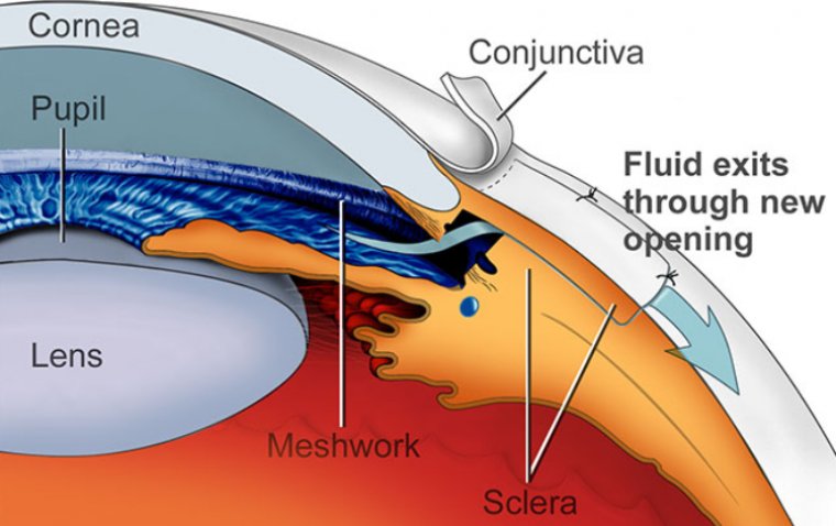
● Sclera
The sclera is the tough, white outer layer of the eye. It provides protection and structure to the eye. The sclera is made up of collagen and elastic fibers and is the thickest layer of the eye. The sclera is continuous with the cornea at the front of the eye and with the optic nerve at the back of the eye.
● Fovea
The fovea is a small, central area of the retina that is responsible for providing detailed, high-resolution vision. It is located at the center of the macula, which is the oval-shaped area at the center of the retina. The fovea contains a high concentration of cone cells, which are responsible for detecting color and fine details.
● External Eye Muscles
The external eye muscles are a group of six muscles that are responsible for controlling the movement of the eye. These muscles are attached to the outer surface of the eye and work together to move the eye in different directions.
The six external eye muscles are:
1. Lateral rectus muscle: This muscle is located on the outer side of the eye and is responsible for moving the eye outward, away from the nose.
2. Medial rectus muscle: This muscle is located on the inner side of the eye and is responsible for moving the eye inward, towards the nose.
3. Superior rectus muscle: This muscle is located above the eye and is responsible for moving the eye upward.
4. Inferior rectus muscle: This muscle is located below the eye and is responsible for moving the eye downward.
5. Superior oblique muscle: This muscle is located in the upper and back part of the eye and is responsible for moving the eye downward and inward.
6. Inferior oblique muscle: This muscle is located in the lower and back part of the eye and is responsible for moving the eye upward and outward.
In conclusion, the anatomy of the eye is a complex and intricate system that works together to help us see. Each part plays a critical role in the process of vision, from the cornea that refracts light to the retina that processes visual information. Understanding the anatomy of the eye can help us appreciate its complexity and importance in our daily lives.
FAQ