(1).jpg)

Researchers Investigate the Effects of Pegcetacoplan on GA Progression
Researchers from the National Institute for Health Research Biomedical Research Centre at Moorfields Eye Hospital and University College London Institute of Ophthalmology, London have conducted a post hoc analysis of data from two phase 3 clinical trials of pegcetacoplan (Syfovre, Apellis Pharmaceuticals). Their findings suggest that pegcetacoplan may delay atrophy of the retinal pigment epithelium (RPE) and photoreceptors in patients with geography atrophy (GA).
Streamlining GA Evaluation with Advanced Imaging Techniques
Spectral-domain optical coherence tomography (SD-OCT) is commonly used to evaluate GA. However, according to the authors, determining changes in GA progression based on the Consensus of Atrophy Meetings (CAM) group requires manual segmentation of OCT volume scans, which is time-consuming, labor-intensive, and limited by interrater variability. To address this, the authors developed a deep-learning based platform that uses CAM-defined OCT features to detect and quantify GA and its components, streamlining the process.
Insights from a Comprehensive Post Hoc Analysis
The post hoc analysis included 11,614 SD-OCT volumes from 936 of the 1,258 participants with GA in the OAKS (NCT03525613) and DERBY (NCT03525600) studies, which were 24-month, multicenter, randomized, double-masked, sham-controlled studies. All participants had been treated with pegcetacoplan, either monthly or every other month, or with sham injection. The primary endpoint was the least-squares mean change from baseline in the area of RPE and outer retinal atrophy at 24 months.
Pegcetacoplan's Impact on GA Progression
Among the participants, pegcetacoplan treatment was associated with reduced growth rates of SD-OCT biomarkers for GA for up to 24 months compared to sham treatment. Reductions in the least-squares mean change from baseline values of RPE and outer retinal atrophy area were detectable at every time point from 3 through 24 months. The association was more pronounced with more frequent dosing. Stronger associations were observed in the parafoveal and perifoveal regions for both monthly and every other month dosing.
"These findings offer additional insight into the potential effects of pegcetacoplan on the development of GA, including potential effects on the retinal pigment epithelium and photoreceptors," Fu and associates concluded.
About Geographic Atrophy
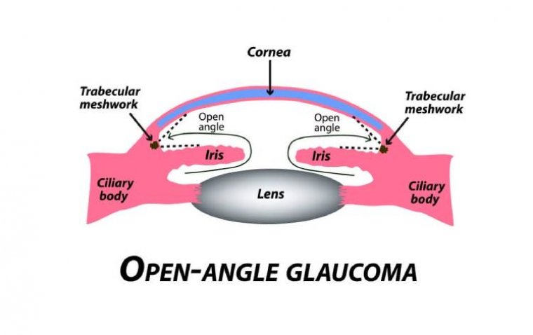
Geographic atrophy (GA) is a progressive and irreversible form of advanced age-related macular degeneration (AMD), a leading cause of blindness in the elderly. GA is characterized by the gradual loss of retinal pigment epithelium (RPE) cells, which are essential for maintaining the health and function of the retina. As RPE cells deteriorate, regions of the macula become thinned and depigmented, resulting in the formation of distinct patches of atrophy that resemble geographic shapes hence the name "geographic atrophy."
Reference
Fu DJ, Bagga P, Naik G, et al. Pegcetacoplan treatment and consensus features of geographic atrophy over 24 months. JAMA Ophthalmol. Published online May 9, 2024. doi:10.1001/jamaophthalmol.2024.1269