(1).jpg)
Scientists Develop Simulations to Help Surgeons Choose the Best IOL Option
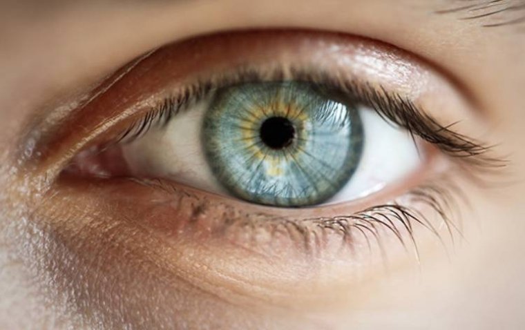
Scientists have developed computer models of patients' eyes to determine the most suitable intraocular lenses and to offer patients a visual simulation of their expected vision with these lenses.
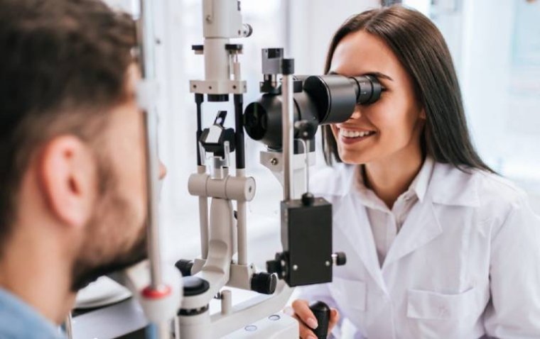
Although millions of individuals have undergone LASIK eye surgery since it was first commercially available in 1989, some patients develop cataracts later in life, necessitating the implantation of new corrective lenses. As the range of intraocular lens options continues to grow, researchers have introduced computational simulations to helpp patients and surgeons in selecting the most suitable options.
In a study featured in the Journal of Cataracts & Refractive Surgery, scientists from the University of Rochester constructed computational eye models that incorporated the corneas of patients who had previously undergone LASIK surgery. These models were used to evaluate the performance of standard intraocular lenses as well as lenses designed to enhance depth of focus in surgically treated eyes.
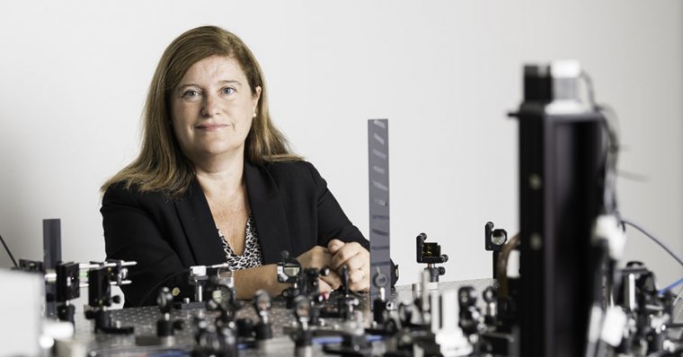
Susana Marcos, the David R. Williams Director of the Center for Visual Science and the Nicholas George Professor of Optics and Ophthalmology at Rochester, emphasized that these computational models, which utilize anatomical data from the patient's eye, provide surgeons with crucial guidance regarding the expected optical quality following surgery.
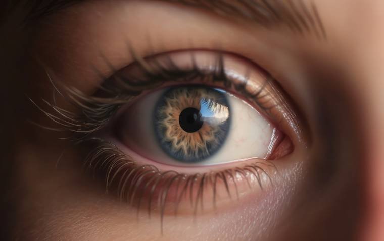
“Currently the only pre-operative data used to select the lens is essentially the length and curvature of the cornea,” says Marcos, a coauthor of the study. “This new technology allows us to reconstruct the eye in three dimensions, providing us the entire topography of the cornea and crystalline lens, where the intraocular lens is implanted. When you have all this three-dimensional information, you’re in a much better position to select the lens that will produce the best image at the retinal plane.”
The Future of OCT
Marcos and her team, in collaboration with the Center for Visual Science, Rochester's Flaum Eye Institute, and Goergen Institute for Data Science, are currently engaged in an extensive study. This study aims to employ their proprietary optical coherence tomography quantification tools to quantify eye images in three dimensions, uncovering overarching patterns and trends. Utilizing machine-learning algorithms, they seek to establish connections between pre- and post-operation data, which will yield valuable parameters for optimizing outcomes.
Furthermore, they have also developed technology that enables patients to visualize the visual effects of various lens options for themselves.
“What we see is not strictly the image that is project on the retina,” says Marcos. “There is all the visual processing and perception that comes in. When surgeons are planning the surgery, it is very difficult for them to convey to the patients how they are going to see. A computational, personalized eye model tells which lens is the best fit for the patient’s eye anatomy, but patients want to see for themselves.”
Using an optical bench, the researchers use technology initially designed for astronomy, such as adaptive optics mirrors and spatial light modulators. They employ these tools to manipulate the optics of the eye in a manner similar to an intraocular lens. This innovative approach enables Marcos and her collaborators to conduct essential experiments and collaborate with industry partners to assess novel products. Marcos has also contributed to the development of a commercial headset version of this equipment, known as SimVis Gekko, which allows patients to experience the world around them as if they had undergone the surgical procedure.
In addition to their research on cataract treatment techniques, the team is applying their methodologies to investigate other significant eye conditions, including presbyopia and myopia.