(1).jpg)

New Study Reveals Rare Cells as Possible Missing Link in Color Perception
In a study published in the Journal of Neuroscience, researchers at the University of Rochester have made significant strides in understanding how humans perceive color. Utilizing adaptive optics, the team has identified rare retinal ganglion cells (RGCs) that could reshape our understanding of color vision.
The Role of Cone Photoreceptors
The retina contains three types of cone photoreceptors sensitive to different wavelengths of light—short, medium, or long. These cones relay information to the central nervous system via retinal ganglion cells. While most RGCs adhere to the well-established "cardinal directions" of color detection, outlined by David Williams, the William G. Allyn Professor of Medical Optics, in the 1980s, discrepancies remain in our understanding of how the eye perceives colors versus how colors are represented.
Discovery of Non-Cardinal RGCs
The recent discovery by the Rochester team, which includes members from the Center for Visual Science, the Institute of Optics, and the Flaum Eye Institute, centers on non-cardinal RGCs located in the fovea. These cells potentially elucidate the complex process behind the human perception of basic colors like red, green, blue, and yellow.
"We don't really know anything for certain yet about these cells other than that they exist," explains Sara Patterson, a postdoctoral researcher who led the study. "There's so much more that we have to learn about how their response properties operate, but they're a compelling option as a missing link in how our retina processes color."
Adaptive Optics: Enhancing Ophthalmic Research
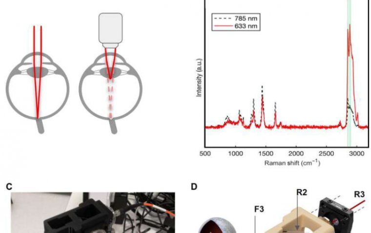
The study also highlighted the use of adaptive optics, a technique originally developed for astronomy to reduce atmospheric blurring in images. This technology was adapted for ophthalmology in the 1990s by Williams and his team. It involves a deformable mirror that corrects light distortion, allowing for a clearer view of the eye's interior and its cells.
"The optics of the eye's lens are imperfect, which reduces the resolution achievable with standard ophthalmoscopes," Patterson says. "Adaptive optics helps to correct these aberrations and provides a crystal-clear view of the retinal ganglion cells, which are the sole source of visual information to the brain."
Understanding these complex cellular mechanisms is not just an academic pursuit. It holds practical implications for improving vision restoration techniques. "Humans have more than 20 ganglion cells, and our models of human vision are only based on three," Patterson remarks. "If we understood the full range of activities of these cells, we could enhance the functionality of retinal prosthetics to better mimic natural visual processing."
This research not only offers insights into the biological underpinnings of sight but also paves the way for future innovations in visual rehabilitation technologies.
Reference
Tyler Godat et al, Cone-Opponent Ganglion Cells in the Primate Fovea Tuned to Non-Cardinal Color Directions, The Journal of Neuroscience (2024). DOI: 10.1523/JNEUROSCI.1738-23.2024