(1).jpg)
Optical coherence tomography (OCT) – Optimizing Clinical Care
Optical coherence tomography (OCT) has been a cornerstone tool for many years for the diagnosis and management of conditions involving the posterior segment.
Although continuous hardware and software advances have increased the applications and utility of OCT, multimodal imaging that allows a more comprehensive assessment of pathology may be helpful and even integral in some situations for guiding accurate diagnosis, appropriate treatment, and optimal follow-up.
DRI OCT Triton™ from Topcon Healthcare addresses this need. When released in 2014, Triton was the first combined anterior and posterior swept-source OCT system that also incorporated full colour and high resolution stereo fundus photography, as well as fluorescein angiography and autofluorescence imaging.
.jpg)
In 2015, Triton’s functionality was expanded with the addition of OCT angiography (OCTA). Then, in 2020, OCT image quality was improved with the release of PixelSmart™, Topcon’s new image processing algorithm.
Topcon’s history of staying at the forefront in introducing cutting edge OCT technology explains why Topcon Healthcare continues to be a leading manufacturer in the competitive field of OCT instrumentation.
State-of-the art attributes Triton’s swept-source OCT technology has the advantage of being able to scan through media opacities and offers unparalleled imaging depth and quality.
The OCT module uses a 1050-nm light source with high signal-to-noise ratio provided by swept-source combined with the auto-mosaic panorama facility, allowing Triton to create a montage of images that include both the central macula and the periphery.
(1).jpg)
PixelSmart technology reduces speckle noise, improves contrast, delivers enhanced image quality for all OCT 3D scans (3D wide, 3D macula, 3D disc, and combination scans), and can be applied to existing scans without changing metrics.
A new software upgrade brings improved OCTA imagery and metrics by enhancing objective and quantitative assessment of retinal vasculature. Moreover, efficiency of imaging and data acquisition are optimized by Triton’s ultra-fast scanning speed (100,000 A-scans/sec) and proprietary eye-tracking system (SmartTrack™).
The fast scanning speed helps to reduce artifacts from involuntary eye movements, enabling capture of clear images even in patients with unstable fixation. The likelihood of movement artifacts is also reduced by Triton’s nearly invisible scanning line, assisting patients to concentrate on the fixation target.
These features help to overcome capture challenges that are particularly likely with the youngest patients and geriatric individuals, but can occur with any patient. Reducing the likelihood that rescanning is needed further supports efficiency and clinic workflow.
.jpg)
Experts’ perspective The following cases from leading retina and uveitis specialists illustrate the utility and benefits of multimodal imaging using Triton.
CASE 1: Bilateral Proliferative Diabetic Retinopathy with Macular Edema Following Laser Photocoagulation Luis Arias Barquet, MD, PhD, Head of the Retina Service, University Hospital of Bellvitge, and Aggregate Professor of Ophthalmology, University of Barcelona, Spain, shared a case of a 69-year-old white male with diabetic retinopathy.
The patient has a 10-year history of diabetes for which he has used oral medication and insulin. He had previously been treated for diabetic retinopathy in both eyes with laser photocoagulation, but reported progressive visual loss over the past month.
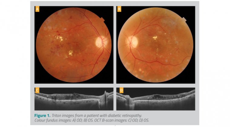
His visual acuity was 20/50 OD and 20/80 OS. Fundus photography in both eyes showed pale optic discs, macular thickening with hard exudates, microaneurysms, and scars from the laser photocoagulation (Figures 1A, B).
On the OCT B-scan images, severe macular edema with large cysts were present OU; the outer retinal layer was relatively preserved OD, but compromised OS (Figures 1C, D). The latter discontinuity in the retinal layers was believed to explain the worse vision OS.
OCTA images, OD and OS, are shown in Figures 2A and 2B, respectively, and the findings are similar for the two eyes. Superficial plexus evaluation showed enlargement of the foveal avascular zone with capillary dilation, microaneurysm, and areas of peripheral ischemia (top rows, orange frames).
Dark areas in the deep plexus correspond to the inter-retinal cysts (top rows, green frames). The en face OCT images (bottom rows, middle images) help to distinguish the cysts from ischemia. Dr. Arias Barquet said, “The collective findings from comprehensive imaging are essential for planning optimal management of patients with diabetic retinopathy.
This case demonstrates the clinical advantages of utilizing Triton multimodal imaging.” He explained, “Fundus photography with Triton allows visualization of macular thickening, hard exudates, and blood.
The OCT B-scans permit accurate assessment of the severity of the macular edema and support evaluation of the integrity of the outer retinal layer that may inform visual prognosis. The OCTA and en face imaging enable the identification of ischemia, vascular alterations, and cysts.”
CASE 2: Wet Age-Related Macular Degeneration Heloisa Nascimento, MD, PhD, Department of Ophthalmology, Federal University of Sao Paulo, Paulista Medical School – UNIFESP/EPM, Brazil, shared two cases.
The first case involved an 80-year-old female patient who presented with wet age-related macular degeneration. Triton multimodal imaging captured a colour fundus image (Figure 3A) and OCT images (Figure 3B) simultaneously with a single acquisition.
Commenting on the benefit of PixelSmart, Dr Nascimento said, “Having good images with a high-quality scan allows for better evaluation of retinal and choroidal alterations.
This is especially important when imaging patients who are not cooperative during the exam or have poor fixation, making it difficult to precisely position a line or radial high-quality scan at the site of the abnormality.”
.jpg)
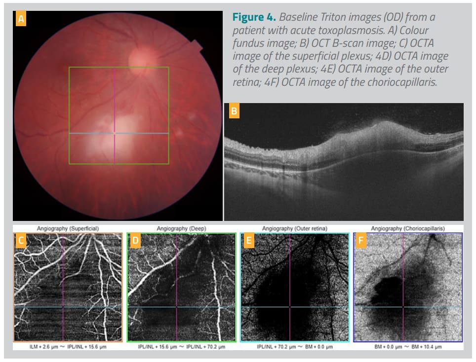
CASE 3: Acute Toxoplasmosis Dr. Nascimento ’s second case involved a 20-year-old female diagnosed with acute toxoplasmosis. The patient presented with complaints of floaters, photophobia, and ocular pain in her right eye.
The Triton colour fundus image showed a white focal retinitis with overlying vitreous inflammation, vasculitis, and hemorrhages (Figure 4A). Even though there was significant vitreous opacity, the OCT image revealed posterior vitreous inflammation with intraretinal alterations and focal choroidal thickening (Figure 4B).
Using Triton’s OCTA, neovascularization was ruled out, and focal areas of non-perfusion in the superior plexus were seen (Figures 4C-F). Those findings, together with the patient history, led to the diagnosis of acute toxoplasmosis. The patient was treated with oral antibiotics and steroids.
Repeat imaging with Triton after 5 months showed the toxoplasmosis lesion had become well delineated and had more pigment at its borders, the vasculitis had resolved, and vitreous inflammation was also improved (Figure 5A).
OCT showed reduction in inflammation, lesion size, and choroidal thickening; partial vitreous detachment with posterior vitreous thickening was also visible (Figure 5B). OCTA showed improvement in the retinal vascularization that was adjacent to the lesion (Figures 5C-F).
Dr. Nascimento noted that multimodal imaging utilizing Triton guides her clinical decisions for patients with uveitis, both for establishing the underlying etiology and determining appropriate treatment.
“Uveitis has a broad range of infectious and noninfectious causes, making differential diagnosis important and sometimes challenging. In cases of posterior uveitis, Triton’s swept-source OCT helps me to visualize whether the retina and/or choroid is involved and to distinguish between causative infectious agents, such as herpesvirus, toxoplasmosis, or syphilis.
For patients with uveitis-related cystoid macular edema, imaging with Triton helps support my plan for treatment and subsequent follow-up,” Dr. Nascimento.
Staying at the top not only describes Topcon Healthcare’s top-ranking market share in the field of OCT technology, but also the company’s dedication to perpetual innovation, leading to new tools and enhancements that support practice workflow efficiency for clinicians and their delivery of optimal patient care.
The introduction of the multimodal Triton OCT in 2014, the addition of OCTA in 2015, and the release of PixelSmart technology in 2020 reflect Topcon Healthcare’s dedication to meeting the needs of eye care professionals and the patients they serve.