What You Need to Know About Dermoid Cyst in the Eye
What Is Dermoid Cyst in the Eye?
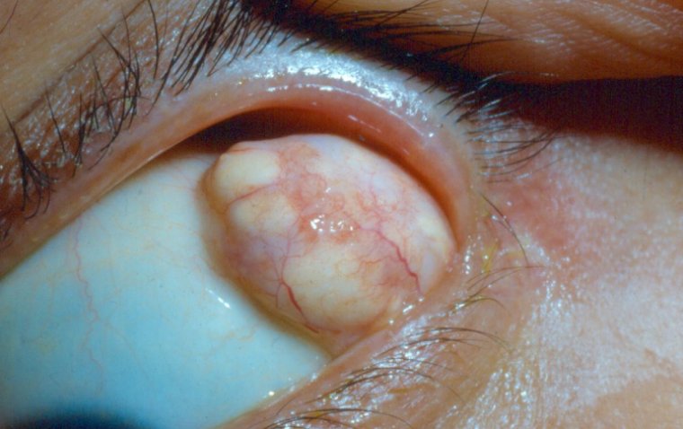
A dermoid cyst in the eye is a benign, congenital growth that can appear on or around the ocular structures. These cysts are unique because they contain a range of tissue types commonly found in the skin layers, such as hair follicles, sweat glands, and sometimes even teeth or cartilage. Unlike regular cysts, which are filled with fluid, dermoid cysts have a more complex internal composition owing to the variety of tissues they can contain. They typically manifest near the eye's surface but can sometimes extend deeper into orbital tissues.
What Causes Dermoid Cysts?
Dermoid cysts are generally thought to have an embryonic origin, arising from misplaced tissue during fetal development. Specifically, during the formation of the skin and associated structures, certain cells may get trapped in layers where they don't normally belong. Over time, these misplaced cells can lead to the growth of a dermoid cyst.
In the context of ocular dermoid cysts, these formations can occur in various parts of the eye, most commonly affecting the outer layer of the eye, such as the conjunctiva and cornea. However, they can sometimes be found deeper within the orbital structure, including within the eyelid or near the optic nerve. The variety of possible locations is consistent with the notion that dermoid cysts result from developmental irregularities.
Signs and Symptoms of Dermoid Cysts
Dermoid cysts in the eye may present with a range of symptoms depending on their size, location, and rate of growth. The most common and evident symptom is the presence of a visible lump or mass on the eye, often on the surface layers like the conjunctiva or cornea. This lump may be flesh-colored, yellowish, or have a whitish appearance.
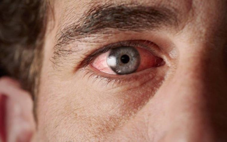
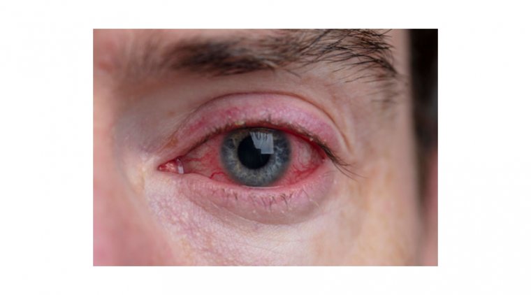
Discomfort or a gritty sensation in the eye could also be a symptom, particularly if the cyst is large enough to interfere with the natural movements of the eye or eyelids. In some cases, redness or inflammation may occur around the area where the cyst has formed. If the cyst is located in a critical area that affects the pathway of light into the eye, it could even interfere with vision, causing blurred or obstructed sight.
Additionally, it's worth noting that the appearance and symptoms of a dermoid cyst can change over time. Depending on factors such as growth rate and potential for irritation, the cyst might become more noticeable or symptomatic as time progresses. Therefore, ongoing monitoring is essential for appropriate management.

How to Diagnose Dermoid Cysts
Diagnosing a dermoid cyst in the eye typically begins with a thorough clinical examination. Your healthcare provider may conduct a comprehensive eye examination, inspecting the cyst visually and palpating it to ascertain its texture, size, and location. The eye may also be examined under a slit lamp, a special microscope used to evaluate the different parts of the eye in greater detail.
Advanced imaging techniques can provide a more comprehensive understanding of the cyst and its relationship to surrounding structures. Ultrasound may be employed to visualize the cyst and assess its density, helping to differentiate it from other eye abnormalities. Magnetic Resonance Imaging (MRI) can offer even more detail and is particularly useful if the cyst is deep or located close to critical structures within the eye.
In some cases, a biopsy might be considered, especially if there's uncertainty about the nature of the lump. This involves taking a small tissue sample from the cyst for histopathological examination to confirm its benign nature and rule out malignancy or other conditions.
Treatment Options for Dermoid Cysts
The treatment approach for dermoid cysts depends on various factors such as the cyst's size, location, and potential impact on vision. Here are some of the options:
1. Surgical Removal
Surgical excision is often the primary treatment for dermoid cysts, especially those that are large, visually prominent, or causing discomfort. The surgery aims to remove the cyst completely to prevent recurrence. Surgical techniques can vary depending on the cyst's size and location. For example, if the cyst is superficial and located on the conjunctiva, a relatively simple surgical excision might suffice. However, if the cyst is deep or near critical eye structures, a more complex surgical approach may be needed.
The ultimate goal of surgery is to remove the cyst while minimizing any cosmetic or functional impairment. The surgeon will generally aim for minimal scarring and optimal visual outcomes. This is particularly crucial if the cyst is located near structures critical to vision, such as the cornea or optic nerve.
2. Non-Surgical Approaches
In some cases—usually when the cysts are small and not causing any symptoms—watchful waiting might be an option. These cysts may be monitored through regular eye examinations and imaging to ensure they're not growing or causing complications. However, surgical removal is typically recommended for definitive treatment.
The choice of treatment should be individualized, taking into account factors like patient age, general health, and the specific characteristics of the cyst. Consulting an ophthalmologist who specializes in ocular tumors or orbital diseases is often recommended to get a comprehensive evaluation and personalized treatment plan.
Potential Complications
● Cyst Growth
One of the significant concerns with dermoid cysts is their potential to grow over time. While these cysts are generally slow-growing, an increase in size could exacerbate symptoms and complicate surgical removal. Larger cysts are more likely to impact vision and could pose greater risks during surgery.
● Infection
Though relatively rare, dermoid cysts can become infected. An infected cyst can cause severe discomfort, redness, and may require immediate medical intervention, such as drainage and antibiotic treatment, to control the spread of infection.
● Disruption of Eye Function
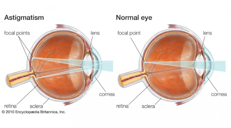
Depending on their location, larger cysts could potentially interfere with essential eye functions like tear production or eye movement. They might also induce astigmatism if they exert pressure on the cornea.
Lifestyle and Aftercare
Post-Surgical Care
After the surgical removal of a dermoid cyst, it's crucial to adhere to post-surgical care guidelines to ensure optimal healing and minimize complications. These guidelines often include using prescribed antibiotic and anti-inflammatory eye drops to prevent infection and control inflammation. Additionally, you may be advised to use a protective eye shield, especially during sleep, to avoid any accidental rubbing or pressure on the surgical site.
Recovery Period
The recovery period can vary depending on the complexity of the surgery and your general health status. During this time, you may experience some discomfort, redness, or temporary vision changes, which are typically normal and should gradually improve. You should have a follow-up appointment with your ophthalmologist to assess the healing process and make any necessary adjustments to your treatment plan.
Importance of Ophthalmologist's Instructions
Following your ophthalmologist's instructions is of the utmost importance for achieving the best possible outcome. This includes taking all medications as prescribed, avoiding strenuous activities that may strain the eyes, and returning for scheduled follow-up examinations. Non-adherence to post-surgical instructions could lead to complications, such as infection or inadequate healing, which could potentially impact your long-term vision and eye health.
Is It Possible to Prevent Dermoid Cysts?
Congenital Nature of Dermoid Cysts
It's important to understand that dermoid cysts are usually congenital, meaning they are present from birth. These cysts arise from abnormal tissue placement during embryonic development, making them inherently non-preventable through lifestyle changes or medical interventions after birth.
Limitations on Prevention Strategies
Because dermoid cysts are typically present from birth, traditional prevention strategies are not applicable. There's currently no known method for preventing the formation of these cysts during pregnancy, and they are not thought to be linked to any specific environmental factors, lifestyle choices, or medical conditions that could be modified for prevention.
Prenatal Care
While the prevention of dermoid cysts isn't possible, appropriate prenatal care, including regular medical check-ups and imaging studies, can sometimes detect anomalies early on. Although this doesn't prevent the condition, early detection could offer more options for management and prepare parents and healthcare providers for postnatal care.
Summary
In this article, we've explored the complex topic of dermoid cysts in the eye. These cysts are congenital formations that occur due to abnormal tissue placement during embryonic development. They can appear in various parts of the eye and come with their own set of symptoms, ranging from visible lumps to potential discomfort and redness.
Accurate diagnosis is critical and typically involves clinical examination, supplemented by imaging techniques like ultrasound or MRI, and occasionally, biopsy. The differentiation of dermoid cysts from other eye conditions is crucial for the appropriate treatment.
Treatment usually hinges on several factors including the size, location, and impact of the cyst on vision. Surgical removal is a common approach, designed to balance both cosmetic and functional outcomes.
We also delved into the possible complications of untreated or poorly managed dermoid cysts, such as cyst growth and infection, particularly stressing the importance of regular monitoring in pediatric cases. Post-surgical aftercare is another significant aspect of effective management.
Though dermoid cysts are congenital and not preventable, we touched upon the value of early detection through prenatal care. Given the complexity of diagnosing and treating dermoid cysts, it is crucial to consult with healthcare professionals for an accurate diagnosis and individualized treatment plan. Early detection and appropriate management are key to mitigating complications and improving outcomes.
(1).jpg)
