(1).jpg)
New Type of Neuron Discovered to Guide Formation of Retinal Blood Vessel Lattice
Scientists at UC San Francisco have identified a novel type of neuron that plays a crucial role in guiding the formation of the intricate lattice of blood vessels in the retina. This lattice is essential for nourishing the cells responsible for vision.
The findings, published in the journal Cell, offer insights that could potentially lead to new therapeutic avenues for diseases linked to impaired blood flow in the eyes and brain.
Perivascular Neurons at the Forefront of Retinal Vessel Formation
Dr. Xin Duan, an associate professor of ophthalmology and senior author of the study, expressed the significance of the discovery, stating, "This is the first time anyone has seen retinal neurons using direct contact with blood vessels as a way of guiding them to form these precise 3D lattices." He further added, "This brings us closer to the possibility of repairing them when they're damaged or rerouting them when they weren't built right in the first place."
Unveiling the PIEZO2 Connection
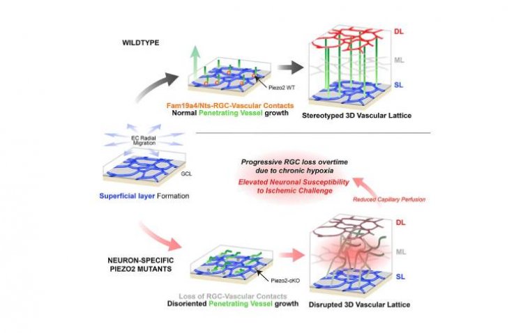
The researchers focused their investigations on newborn mice, whose eyes undergo significant development in the weeks following birth. Dr. Kenichi Toma labeled retinal neurons adjacent to blood vessels with a glowing protein, enabling the observation of the lattice formation process. Through their work, they identified a specific subset of neurons, termed perivascular neurons, which interact with and envelop growing blood vessels, orchestrating their arrangement into the lattice structure. These perivascular neurons produce a protein known as PIEZO2, allowing them to sense contact with neighboring cells.
Notably, mice lacking the ability to produce PIEZO2 exhibited an inability to maintain contact with blood vessels, resulting in a disorganized growth pattern that impeded proper blood flow. This disruption led to oxygen deprivation in surrounding nerve cells, rendering the mutant mice more susceptible to stroke-like injuries.
The Crucial Role of Cerebellar Lattice Structure
Expanding the scope of their research, Dr. Duan and his team discovered that perivascular neurons also play a crucial role in guiding the formation of blood vessel networks in the cerebellum, a region of the brain associated with coordination and sensory perception. Dr. Toma emphasized the potential implications of their findings, suggesting that damage to this lattice structure could contribute to various neurodegenerative diseases.
Collaborating with developmental biologist Dr. Arnold Kriegstein, the researchers confirmed the existence of perivascular retinal neurons in humans, further underscoring the relevance of their discoveries beyond animal models.
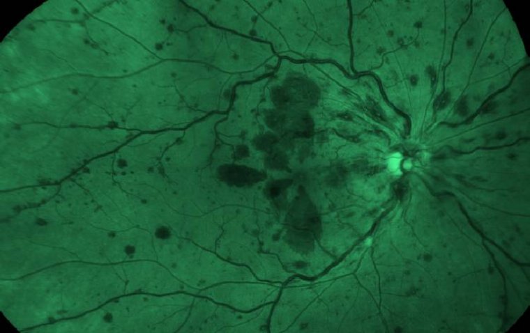
Central to their investigation was the utilization of advanced imaging techniques, including multiphoton microscopy developed by Dr. Tyson Kim. This cutting-edge approach allowed for the generation of detailed 3D images of retinal blood networks, offering unprecedented insights into the formation and breakdown of the lattice structure.
Implications for Treating Neurodegenerative Diseases
Dr. Kim highlighted the collaborative nature of their research, stating, "It was really a confluence of what we're each passionate about." The synergistic efforts of the multidisciplinary team paved the way for a deeper understanding of the intricate relationship between the vascular and nervous systems.
The implications of these discoveries extend beyond fundamental neuroscience, potentially offering new avenues for treating neurodegenerative diseases by safeguarding the intricate networks of blood vessels that support neuronal function. As Dr. Duan aptly summarized, "There are lots of people trying to understand the ways we can grow neurons. But how in the world do we grow the intricate networks of blood vessels required to support them? That's the question we're trying to answer."
Reference
Kenichi Toma et al, Perivascular neurons instruct 3D vascular lattice formation via neurovascular contact, Cell (2024). DOI: 10.1016/j.cell.2024.04.010