(1).jpg)

Researchers Introduce Novel Imaging Method for Early Diagnosis of Eye Diseases
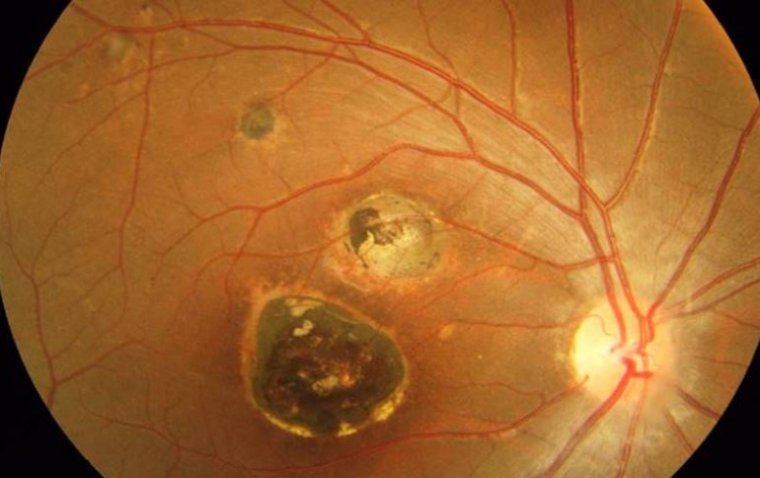
Optical coherence tomography (OCT) stands as one of the most foundational and accurate tests for diagnosing eye diseases. While it excels in a detailed examination of individual eye structures, its effectiveness diminishes when applied to the early detection of subtle pathological changes.
In a bid to overcome this challenge, researchers at ICTER have introduced a novel imaging method derived from OCT. This innovation has led to the development of spatio-temporal optical coherence tomography (STOC-T), an even more advanced approach that mitigates noise and facilitates the acquisition of precise images.
STOC-T is particularly instrumental in diagnosing early-stage disease changes and has a notable application known as optoretinography (ORG).
According to the ICTER news release, this solution marks a crucial stride in advancing ocular disease diagnostics. Unlike traditional OCT, which scans the eye with coherent light, STOC-T employs several hundred different laser patterns to swiftly illuminate the retina within nanoseconds. This captures instantaneous reactions, enabling the computational analysis of extensive datasets. Consequently, doctors gain access to highly sophisticated and precise information about the eye's condition, significantly enhancing the visualization of retina and choroid images. This level of detail was previously unattainable.
Olaf Strauss, PhD, an experimental biologist at Charité - Universitätsmedizin Berlin, is actively engaged in research focused on identifying methods for treating blindness.
“Eye function is more critical than structure because often in the course of a disease, we first observe changes in function preceding changes in structure,” Strauss said in the news release. “Therefore, highly sensitive measurements of eye function are crucial for monitoring and detecting pathological changes in tissue.”
According to the center, this technology is set to revolutionize the diagnosis of eye diseases, enabling ophthalmologists to work much faster and more efficiently. Patient examinations will now take just one-hundredth of a second, a remarkable improvement from the several minutes required in current OCT examinations. An ultra-fast camera, with the ability to capture 100,000 frames per second, transmits extensive data to a computer, allowing for the observation of receptors' responses to light.
ICTER highlighted that their software processes this data, generating images comparable to what a microscope provides. At present, ICTER is actively researching specific receptor movements linked to various diseases. This breakthrough will enable swift and highly precise diagnoses of numerous eye conditions, as well as post-therapy monitoring.
Wojtkowski emphasized in the news release that early diagnosis of these conditions could potentially reduce the harmful effects in approximately 90% of cases.
“By employing the STOC-T research method, we provide the opportunity for in-vivo studies of pharmacological therapies, supplying essential information about the quality and efficacy of proposed eye disease treatments," Wojtkowski said in the ICTER news release.
Furthermore, as highlighted by the researchers in the release, the adoption of STOC-T technology may pave the way for the emergence of a clinical research market focused on cutting-edge eye therapies, such as gene therapies. What's more, the compact and portable nature of STOC-T diagnostic equipment positions it as a fitting addition to any ophthalmology clinic once it becomes commercially available.