(1).jpg)

AAO 2024: Several Imaging Modalities Effective in Geographic Atrophy Diagnosis
At the 2024 American Academy of Ophthalmology (AAO) meeting in Chicago, Dr. SriniVas R. Sadda discussed the evolving landscape of imaging technologies for diagnosing geographic atrophy (GA). While traditional methods like flash color fundus photography have been the gold standard for many years, newer modalities are proving more effective in diagnosing and monitoring this progressive retinal condition.
Challenges with Traditional Color Fundus Photography
Flash color fundus photography has long been the primary method for diagnosing GA, primarily relying on identifying sharply demarcated regions with depigmentation and increased visibility of choroidal blood vessels. According to Dr. Sadda, this approach has been effective for many studies but presents limitations in practice.
“We have a definition for atrophy of a sharply demarcated region with depigmentation and increased visibility of choroidal blood vessels,” Sadda explained. “That served us very well for a number of different studies. But the problem is this requires good stereopsis, and sometimes we don’t have sufficient contrast.”
This issue makes traditional fundus photography less practical for clinical trials and certain patients, particularly when precise contrast is required for accurate diagnosis.
Advances in Imaging Modalities for GA
Newer imaging technologies are addressing the challenges of traditional methods. One such tool is blue-light fundus autofluorescence, which has gained prominence in clinical trials for its ability to provide sharper contrast in imaging. However, as Dr. Sadda noted, blue light can be uncomfortable for some patients, and repeated exposure may have toxic effects in individuals with atrophic macular disease.
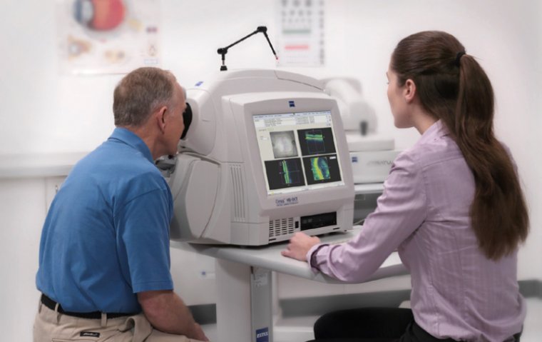
Retina imaging has now advanced into the era of confocal color imaging, green fundus autofluorescence, and optical coherence tomography (OCT). These modalities have proven effective not only in diagnosing GA but also in tracking its progression over time.
Microperimetry for Functional Progression
While not the optimal choice for initial diagnosis, microperimetry plays a crucial role in tracking functional progression in GA patients. Dr. Sadda emphasized its usefulness in evaluating how the disease affects vision over time, making it an important tool for long-term management.
The Future of GA Diagnosis and Management
As more therapies for geographic atrophy become available, technological advancements will continue to refine how GA is diagnosed and managed. Dr. Sadda highlighted the role of artificial intelligence (AI) in this new era, specifically pointing to automatic quantification tools enabled by AI.
"Automatic quantification enabled by AI tools is really going to make a difference in this new era,” he said. These tools will not only streamline the diagnostic process but also enhance the ability to track disease progression, ultimately improving patient care.
Geographic atrophy, a complex and progressive retinal disease, requires precise and reliable diagnostic tools. The evolution of imaging modalities is paving the way for more effective management and treatment of the condition.
Sources: