(1).jpg)

Eye Ultrasound Proves Effective for Pediatric Brain Shunt Failures
Researchers have unveiled promising findings at the Pediatric Academic Societies (PAS) 2024 Meeting in Toronto, indicating that an eye ultrasound could swiftly and safely diagnose brain drainage tube failures in children at emergency departments.
This innovative approach primarily targets children with ventricular shunts—thin, plastic tubes implanted surgically to drain excess fluid and alleviate brain pressure due to hydrocephalus, which can stem from brain bleeds, tumors, or other causes.
The Problem with Current Diagnostic Methods
Historically, nearly 30% of these shunts fail within the first two years of placement due to breaks, displacements, or blockages, with an additional 5% failing annually thereafter. Children presenting symptoms of potential shunt failure, such as headaches, vomiting, and fatigue, often undergo numerous computed tomography (CT) and magnetic resonance imaging (MRI) scans annually. These procedures not only expose them to significant radiation and sedation risks but also present challenges in the timely and accurate diagnosis of shunt failures.
How Eye Ultrasound Can Help
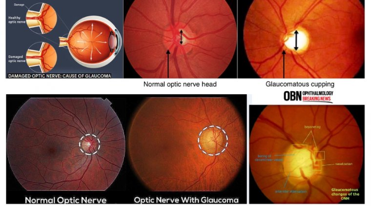
The study introduces a critical advancement in this area by using eye ultrasounds to measure the swelling of the optic nerve sheath, which tends to swell due to fluid backup when shunts fail. By comparing the optic nerve's diameter during symptomatic periods to its size when the child is well, physicians can effectively assess whether a shunt is blocked.
Dr. Adrienne L. Davis, MD, MSc, FRCPC, the pediatric emergency medicine research director at The Hospital for Sick Children (SickKids) in Toronto and the presenting author of the study, emphasized the benefits of this method. "The research team is interested in finding ways to lessen radiation exposure and expedite diagnosing shunt failure in the emergency department," she said. Davis further explained that the study leverages a patient’s previous healthy baseline for comparison, tailoring the diagnostic process to each individual’s unique circumstances and tolerance for brain pressure variations.
Future Directions and Validation
The research was conducted on 76 pairs of eye ultrasounds from nearly 60 children who visited the emergency department of the Toronto hospital with potential shunt failures. While the initial results are promising, the researchers noted that further validation is required across a larger cohort of North American children to confirm the effectiveness and safety of this diagnostic approach.
Reference
Abstract: Change in Optic nerve sheath diameter (ONSD) and Optic disc elevation (ODE) in predicting Shunt failure in the Emergency department