(1).jpg)
Eye Exams May Offer Early Detection Pathway for Alzheimer’s Disease
Routine eye exams could soon serve as a powerful diagnostic tool for detecting Alzheimer’s disease and other forms of dementia, potentially decades before clinical symptoms appear. This is the key finding from a new study conducted by researchers at The Jackson Laboratory (JAX), published in Alzheimer’s & Dementia.
Retinal Vascular Changes Linked to Alzheimer’s Risk
The research highlights abnormal changes in the tiny blood vessels of the retina in mice carrying a common genetic mutation known as MTHFR677C>T, found in up to 40% of people. This mutation has previously been associated with increased Alzheimer’s risk.
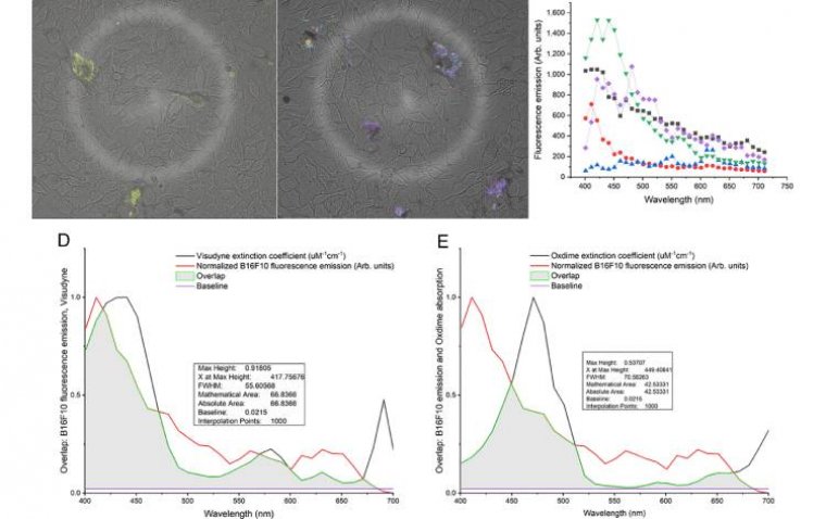
Researchers observed that mice with this mutation showed twisted, narrowed, and swollen retinal vessels, along with reduced vascular branching as early as six months of age. These signs closely resemble vascular changes in the brain associated with poor blood flow and cognitive decline.
“We can see these wavy vessels in the retinas, which can occur in people with dementia,” said Alaina Reagan, neuroscientist at The Jackson Laboratory (JAX).
Reagan noted that such vascular changes likely reflect a systemic issue, not limited to the retina or brain alone, potentially involving hypertension or general vascular dysfunction.
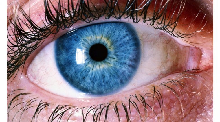
Retina: A Window to the Brain
Because the retina is part of the central nervous system, it shares many structural and functional characteristics with the brain, making it an accessible site for detecting neurological changes.
“Your retina is essentially your brain, but it’s much more accessible because your pupil is just a hole,” Reagan explained. “The neurons, immune cells, and even their responses under disease pressure are quite similar.”
This insight builds upon JAX’s 2022 findings, which identified similar vascular abnormalities in the brains of mice with the same mutation. That earlier study showed fewer vessels in the cortex and reduced cerebral blood flow, providing further support for the retina’s role as a biomarker for neurodegenerative disease.
Molecular Disruptions Mirror Disease Progression
In addition to structural changes, the new study identified protein pattern disruptions in both brain and retinal tissues. These included alterations in cellular energy production, waste protein removal, and vascular support, all of which may contribute to Alzheimer’s progression in individuals with the MTHFR677C>T mutation.
“A lot of these molecular changes are happening in conjunction, which suggests these systems in brain and retinal tissue are working in tandem,” Reagan said.
Clinical Translation and Human Application
To determine whether these retinal vascular changes also occur in human patients, the JAX team is collaborating with clinicians and dementia specialists at Northern Light Acadia Hospital in Bangor, Maine. The goal is to explore how routine eye exams could identify high-risk individuals based on retinal imaging findings.
“Most people over 50 have some kind of vision impairment and get checked annually for prescription changes,” Reagan said.
“Are they more at risk if they have these vascular changes, and is that a point when doctors could start mitigating brain changes? That could be 20 years before cognitive damage becomes noticeable to patients and their families.”
The research underscores the growing recognition of the retina as a non-invasive biomarker for neurodegenerative diseases and raises the possibility that early interventions could begin long before traditional symptoms emerge.
Reference:
Alaina M. Reagan et al, Retinal vascular dysfunction in the Mthfr677C>T mouse model of cerebrovascular disease, Alzheimer's & Dementia (2025). DOI: 10.1002/alz.70501