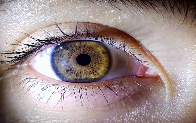
In posterior vitreous detachment (PVD), the gel that fills the eyeball separates from the retina. The retina is a thin layer of nerve tissue that lines the back of the eyeball. It’s responsible for detecting light and turning it into visual images.
Posterior vitreous detachment is a common condition with age. PVD can cause floaters or flashes of light, which often fade over time. PVD isn’t painful or sight-threatening.
The most common symptom of vitreous detachment is a sudden increase in floaters (small dark spots or squiggly lines that float across your vision). When your vitreous detaches, strands of the vitreous often cast new shadows on your retina — and those shadows appear as floaters.
The patient may also notice flashes of light in your side (peripheral) vision. Sometimes, vitreous detachment causes more serious eye problems that need treatment right away. The only way to tell if vitreous detachment has caused a serious eye problem is to get a dilated eye exam.
Managing patients who attend a practice with symptoms of flashes and floaters is a regular occurrence in community optometric practice. It’s often very distressing for patients; symptoms can be quite dramatic and an internet search can indicate imminent blindness from a retinal detachment.
Patients clearly need to be seen swiftly to rule out the possibility of sight-threatening retinal damage. Of course, the majority of patients are able to be reassured with the advice that they should return immediately if anything changes.
A posterior vitreous detachment (PVD) is the most common cause of flashes and floaters in the middle-aged population. Throughout life, the vitreous liquefies and eventually detaches from the retina.
A PVD most commonly occurs between the ages of 55 and 65 years; in general, earlier in myopes and later in hyperopes.
About 50% of the population are symptomatic and so there are numerous patients requiring reassurance that their symptoms are nothing to be alarmed about, and that they don’t need any treatment. This is a normal ageing process.
Of course, there are other causes of flashes and floaters. The differential diagnosis is important to exclude a retinal tear or detachment.
There is an annual incidence of about 10-15 per 100,000 people per year who have a retinal detachment and so whilst this is a rare condition, the sight-threatening nature requires a swift referral for timely treatment to restore and / or retain as much sight as possible.
Modern advancements in surgical procedures have meant that the results are often outstanding.
Taking a detailed patient history
As with many conditions, a detailed history from the patient is vital to being able to exclude many other causes of these symptoms.
For example, migraine auras are another common cause of flashes. More frequently associated with younger patients, and often bilateral, they can be identified with careful questioning by the practitioner.
They may or may not be associated with a headache or migraine. Postural hypotension, another cause of flashes as the patient stands, can also be identified with detailed and specific questioning of the patient.
Alarm bells start to ring when patients describe large and significant changes in their vision – the classic “curtain coming over my vision”, or a dramatic drop in their vision when the macula may be ‘off’.
However, patients are all different and a non-descript history can still elicit a dramatic finding, whilst other patients are extremely disturbed by relatively minor changes to their vitreous.
Clearly a detailed dilated binocular examination of the vitreous and the retina are required. Alwitry et al’s study in 2002 found wide variation in confidence by optometrists to examine and mange a patient presenting with flashes and floaters, with many patients being examined without mydriasis and a lack of understanding about vital signs of tears and detachments.
This was also found in a study using an actor as a model patient. As practice has developed, more practitioners are using a slit-lamp and condensing lens combination routinely to examine patients across the UK in community optometry; indeed, equipment funding and training in Scotland makes this a routine part of the General Ophthalmic Services (GOS) eye examination.
Even without scleral indentation, such an examination will almost certainly identify all patients with tears or full detachments, with those in the very far periphery almost certainly resulting from a traumatic incident.
Asking the patient to move their eyes in eight points of gaze should ensure a view of all but the most peripheral parts of the retina with particular note in the superior temporal quadrant where about 60% of tears occur.
A high magnification slit-lamp is vital to being able to closely examine the vitreous for signs of change. Tobacco dust is a good determinant of a tear with pigment from the retinal pigment epithelium having leaked into the vitreous.
These brown cells in the anterior vitreous are known as ‘Shafer’s sign’. It requires good magnification to see and is best observed by asking the patient to look up and then straight ahead.
Identifying signs of a PVD also gives confidence that this is indeed the cause of the symptoms. Though Weiss’s ring is rarely seen in full, partial remains may be viewed in the vitreous around the disc, near to where the vitreous gel was attached.
Expert opinion informs the National Institute for Health and Care Excellence (NICE) recommendation on referral pathways.
Referral directly to ophthalmology where there are clear signs of retinal detachment or symptoms such as distorted or blurred vision or visual field loss (curtaining or dark shadows) is aimed at GPs or other community care practitioners.
Otherwise refer to a “practitioner competent in the use of slit-lamp examination and indirect ophthalmoscopy,” i.e. a community optometrist, is the advice.
It’s important that patients diagnosed with a PVD are advised to return for another examination if their symptoms return. A period of six to eight weeks appears to be the critical time period over which a partial PVD completely detaches and after which the threat of a retinal tear diminishes.
There is still debate as to whether a follow-up appointment should be booked for the patient. The danger of this strategy is that patients may wait for their appointment rather than returning immediately when new symptoms appear.
Indeed, much of the evidence points to tears developing after an initial review only in patients that have experienced new symptoms.
What is important is to provide the patient with some written information about their condition and what to do if their symptoms should return or worsen. There is some very good information that is available for optometrists to hand to patients.
Examples include the Association of Optometrists, Royal National Institute of Blind People (RNIB) and the College of Optometrists.
Even if a practitioner is very experienced, not all examinations are straightforward. Not all patients have a clear media for example; cataracts or longer standing vitreous changes can make examining the whole retina more difficult. A good dilation is vital, leaving the patient time to fully dilate.
Finding adequate time to see emergency presentations can also be a challenge. Whilst these examinations are usually funded around the country either as part of GOS, or as an enhanced service, there may not be an appointment slot waiting available for patients to be seen right away.
Patients are understandably concerned and stressed, and may have been advised by their GP to be seen urgently. Practitioners can often end up reviewing these patients into their lunchtimes or missing breaks.
Leaving appointment slots available to manage emergencies may be a solution in a large practice, though it might not be practical in a small premise where unfilled slots can impact on the overall revenue of a practice.
It would perhaps be timely to review the management of emergencies in optometric practice and whether staff are given enough time to adequately manage these cases.
As more eye care gets shifted to the optometrist, managing a diary to ensure an appropriate duty of care to the patient, whilst also supporting our professionals, is something that will become an increasing challenge.
In a recent paper, it was identified that managing patients who attend with the symptom of ‘flashes and floaters’ in optometric practice was one area of practice that optometrists in Scotland felt was an area of concern.
Barriers to managing a patient within the community and not referring them on for ophthalmological view included: pressure from the patient to refer them, a general lack of feedback from referrals to ophthalmology and concern about legal action if the patient is not referred and they should have been.
It is perhaps unsurprising that the research showed that optometrists need confidence in their own decision-making to ensure that appropriate referrals are made, and that practitioners who reported finding these patients stressful to manage was a barrier to making the correct referral decision; these practitioners may be over-referring patients.
Referring patients as an emergency where the symptoms are as a result of a retinal tear or detachment is clearly important.
When there is doubt about the diagnosis – when a detachment can’t be ruled out, an urgent referral is still required. There should be no cases where these patients are referred routinely or even for a ‘soon’ appointment.
It is of concern, though perhaps not surprising, that research into managing these patients highlighted a group of practitioners who were possibly over-referring their patients due to their concerns about being sued if they made the wrong decision.
Even the most committed practitioner may have lingering doubts about a patient where the possibility of making an error and having missed a peripheral tear results in the patient losing their eyesight.
Keeping accurate records, conducting a thorough patient history and detailed examination, discussing with the patient the findings and providing them with information and advice to return if things change, constitute good care.
Perhaps, not having any doubts is a sign of arrogance – not being fully aware that errors can be made. We are all human.
A common response to rising demand for healthcare is to extend the role of healthcare professionals.
A good example of this is the developing role of the community optometrist and their requirement to manage more patients within their practices without referring them on to the hospital eye service when ophthalmology care is not required.
Eye care in the community needs to be conducted safely and the care that is provided needs to be of the highest quality. A PVD is a normal process of ageing and has always been widely managed within community optometry.
All too often though, the threshold for referral to accident and emergency has been low; these patients have been referred unnecessarily across the country by many practitioners.
It is vital that these low risk patients are retained within the community as they clearly do not require ophthalmology referral.
Community optometrists have the training, knowledge and equipment to safely manage these patients, to make an accurate diagnosis and to advise rapid re-assessment if symptoms change.