(1).jpg)
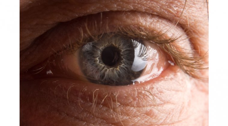
Unveiling Conjunctivochalasis: A Closer Look at the Condition
What Is Conjunctivochalasis?
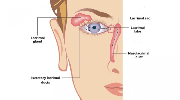
The term conjunctivochalasis was coined by Wendell Hughes in 1942, It means slackening of the conjunctiva which is bound to happen as humans progress in age and the body tissues lose their elasticity. It is a common ocular condition defined as a loose, redundant, non-edematous conjunctiva. Its clinical manifestation and site can differ in patients, however, most frequently it is positioned on the inferior globe. Conjunctivochalasis brings disturbance in the tear film and its drainage pathway.
What Causes Conjunctivochalasis?
The precise etiology of conjunctivochalasis is uncertain, however, it is associated with particular risk factors. Risk factors might activate its early onset, these are stated as under: -
●Eye rubbing, allergic conjunctivitis
●Aging, ocular surface disease
●Dry eyes, contact lens wear
●Hyperopia, pinguecula
●UV exposure
●Eyelid disorders, blepharitis
●Meibomian gland disorder (MGD)
●Ehlers-Danlos Syndrome (EDS)
●Aqueous tear deficiency
●Previous eye surgeries, thyroid disease
Symptoms of Conjunctivochalasis
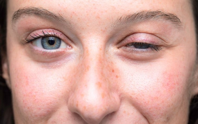
Conjunctivochalasis in its earliest stages is not obvious to either the patient or the ophthalmologist. Some patients with conjunctivochalasis might be entirely symptomless. Before an eye exam, the ophthalmologist will take a brief history of the symptoms experienced by the patient of conjunctivochalasis.
●Insidious beginning of ocular discomfort
●Foreign body sensation or grittiness in eyes
●Dryness
●Excessive tearing or epiphora
●Mild irritation in eyes
●Eyes feeling heavy and sore
●Mucus discharge, nocturnal lagophthalmos
Conjunctivochalasis is often confused with dry eyes, these two are distinguished based on pain that is worsened with a down gaze or blinking solely in conjunctivochalasis and not in dry eyes.
Diagnosis procedures
During a physical clinical examination, using a slit lamp, an ophthalmologist will notice: -
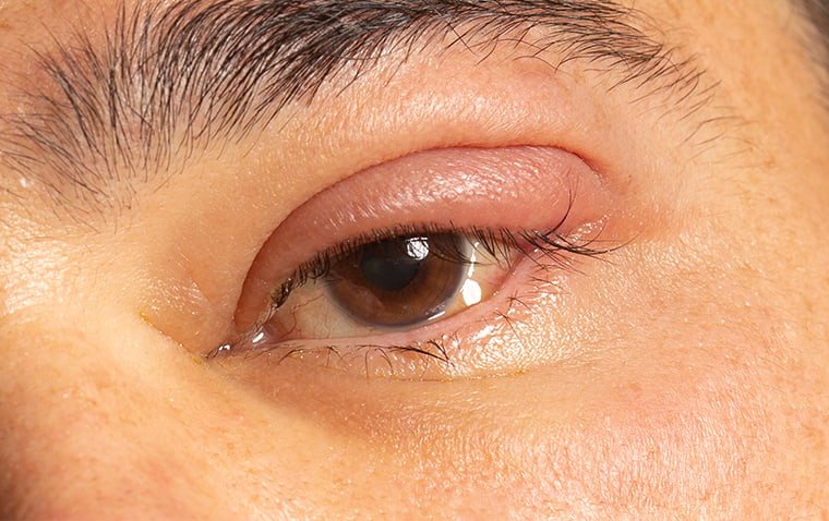
●Redundant conjunctiva overlapping eyelid margin specifically over the inferior temporal region. It could be medial or nasal regions as well.
●Conjunctiva that is effortlessly mobilized by exerting pressure on the eyelid and moving the relaxed and loose conjunctiva up and down.
●In some cases, conjunctival prolapse might occlude the inferior puncta subsequently causing excessive tearing.
An OCT scan can be used to view the anterior segment closely for evaluation of the tear meniscus.
What Are the Treatment Options?
General treatment
Treatment must be customized according to the severity and symptoms of each patient.
In most asymptomatic patients no treatment is required.
Medical therapy
Mild symptoms can be prescribed:
●Ocular lubrication with artificial tears and lubricating ointments to restore the integrity of the tear film.
●Autologous serum teardrops.
●Blinking exercises
●Topical antihistamines
●Topical corticosteroids
●NSAIDs for pain relief
Surgery
Surgery is usually the last option, when every other measure has failed surgery is favored. Multiple surgical options are available to choose from: -
●Conjunctival excision is employed most commonly in it a crescent portion of conjunctiva 5 mm behind the limbus is excised. absorbable sutures or fibrin glue is utilized to close conjunctiva. In other cases, the Amniotic membrane is used to seal in the conjunctival defect.
●Thermal cautery can be done to stiffen the already loose or relaxed conjunctiva.
●Forniceal rebuilding is done with the amniotic membrane to maintain the fornical tear reservoir.
●Argon laser is used to “shrink” the redundant conjunctiva.
●Improving adhesion, to make conjunctiva adhere strongly to the underlying sclera with the aid of scleral fixation.