
The year 2024 has been a remarkable period for advancements in ophthalmology, with groundbreaking in...
read more
The year 2024 has been a pivotal one for ophthalmology, with several novel FDA approvals introducing...
read more
2024 has been a landmark year for breakthroughs in ophthalmology research, showcasing transformative...
read more
In 2024, the ophthalmic industry experienced a transformative year driven by strategic company acqui...
read more
A new analysis published in the British Journal of Ophthalmology reveals that nearly one in three ch...
read more
Lamprey Eye Disease, also known as "Lamprey Disease," is a hoax that has been circulating on the int...
read more
A recent study highlights the potential of ChatGPT-4.0 in accurately answering questions related to ...
read more
Ophthalmology, is facing numerous challenges that impact patient care, innovation, and the overall g...
read more
LASIK surgery has revolutionized the field of vision correction, providing millions of people with c...
read more
Cataract surgery is a safe, effective procedure that improves vision by removing cloudy lenses from ...
read more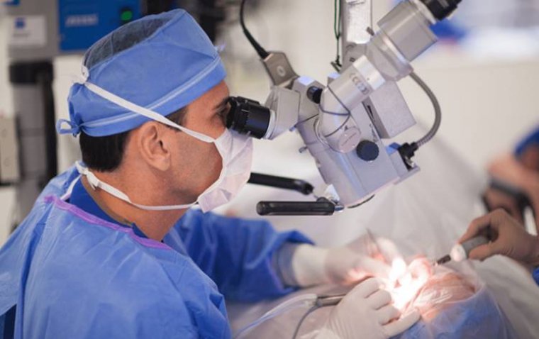
 More
More