(1).jpg)
Nidek Launches the RS-1 Glauvas Optical Coherence Tomography
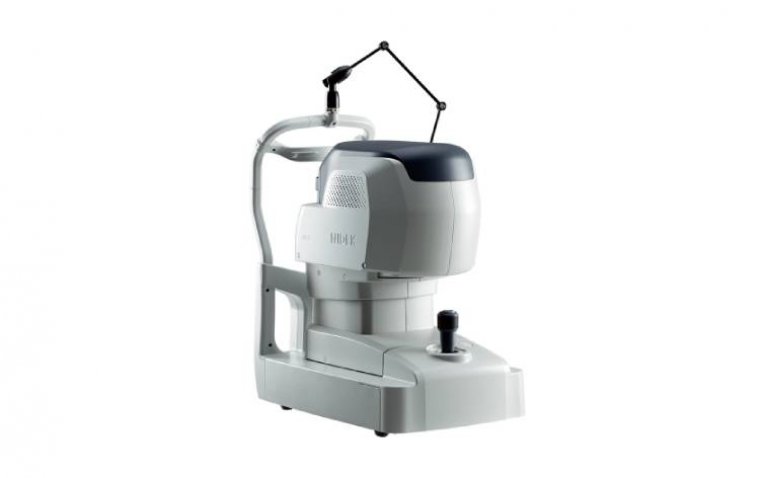
Nidek, a leading provider of ophthalmic equipment, announced the launch of its latest innovation, the RS-1 Glauvas Optical Coherence Tomography (OCT).
The RS-1 Glauvas boasts an impressive scan speed of up to 250kHz, significantly reducing capture time and accelerating workflow. This high-speed imaging not only enhances efficiency but also addresses patient fixation errors, resulting in greater image clarity and improved comfort for the patient, according to Nidek.
"With the capability of wide area imaging, a single B-scan image clearly presents the area from the optic nerve head to the temporal vascular arcade, and the 4.2 mm depth B-scan imaging readily captures the oblate retinal shape of myopic eyes," stated Nidek in a recent news release.
Advanced Analytics and Diagnostic Aid Functions
The device incorporates various advanced analytics functions tailored for the assessment of glaucoma and retinal diseases. Features such as auto alignment, preset scanning patterns, and a user-friendly interface streamline the image capture process, enabling rapid and easy interpretation.
Furthermore, the RS-1 Glauvas utilizes deep learning (DL) segmentation to reduce artifacts and errors in normative databases and thickness maps. This technology, developed in collaboration with Kagoshima University, provides a Structural Normality Map (SN Map) that aids clinicians in detecting minute structural changes at a glance, thereby enhancing diagnostic confidence for early signs of retinal changes.
Addressing Clinical Challenges
One of the key challenges in glaucoma assessment is accurately evaluating patients with axial myopia. To address this, the RS-1 Glauvas incorporates a long axial length normative database and scan width correction, enabling more precise assessments in such cases.
Additionally, the device's DL segmentation reduces false positives and enhances clinic efficiency by minimizing unnecessary follow-up visits. By providing clearer and wider images for assessing chorioretinal microvasculature, the RS-1 Glauvas further contributes to improved diagnostic accuracy and patient care.
Reference:
https://www.nidek-intl.com/news/nidek-launches-the-rs-1-glauvas-optical-coherence-tomography/