(1).jpg)
Researchers Pioneer New OCT Technique for Scleral Structure Analysis
In a study published in the journal JAMA Ophthalmology, researchers at Tokyo Medical and Dental University (TMDU) in Japan have successfully pioneered a new optical coherence tomography (OCT) technique. This innovative method, known as polarization-sensitive OCT (PS-OCT), provides unprecedented insights into the detailed structure of the sclera—the white outer layer of the eyeball.
Motivation Behind the Study
Traditionally, ophthalmologists have faced significant challenges in investigating the finer details of the sclera in living patients. Current techniques have been limited to measuring scleral thickness without revealing the intricate orientation of collagen fibers that compose this crucial eye structure. "The sclera, composed of collagen fibers, plays an important role in protecting the retina, optic nerve, and other neural tissues in the eye. Therefore, abnormalities in the sclera's shape can lead to various complications, including blindness," explains lead author Dr. Kyoko Ohno-Matsui.
Development of PS-OCT
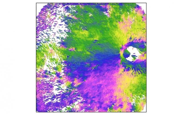
To address these limitations, the TMDU research team developed PS-OCT, a technique where the polarization of light serves as a contrast mechanism. "The sclera exhibits birefringence, an optical property where the refractive index varies with polarization. This property is typically found in fibrous tissues with regularly organized nanostructures, such as the sclera," states senior author Dr. Tae Igarashi-Yokoi. "PS-OCT allows us to not only measure the magnitude of birefringence, indicating fiber density, but also to determine the axis of birefringence, which relates to the orientation of the fiber bundles."
"Thus, in addition to the magnitude of birefringence, which gives us information about the density of fibers, PS-OCT can also show the axis of orientation of the birefringence, which is related to the orientation of the fiber bundles themselves," Dr. Igarashi-Yokoi elaborates.
Research Focus: Highly Myopic Eyes
Using this technique, the team investigated the properties of the collagen fibers in the sclera of patients with highly myopic eyes. They also focused on the link between myopathy and a sometimes-pathological condition known as dome-shaped macula (DSM), in which a specialized area in the retina bulges outwards. Their analysis included 89 highly myopic eyes from a total of 72 patients, mostly adults over 50 years old.
Findings from PS-OCT Imaging
The PS-OCT imaging revealed that the sclera consists of inner and outer layers with distinct structural arrangements. In the inner layer, fibers extend radially from the optic nerve's periphery, while in the outer layer, fibers run perpendicularly. In patients with DSM, the inner layer's fibers were aggregated and thickened, whereas the outer layer's fibers were compressed and thinned.
This innovative use of PS-OCT to visualize fibrous tissue organization in the eye could significantly impact clinical research, diagnostics, and therapeutic strategies. "Recognizing fiber patterns in scleral pathologies such as DSM and staphylomas in myopic eyes can provide valuable insights for developing targeted therapies. Early intervention may help mitigate potential damage to the overlying neural tissue," remarks senior author Dr. Masahiro Yamanari.
Reference
Kyoko Ohno-Matsui et al, Polarization-Sensitive OCT Imaging of Scleral Abnormalities in Eyes With High Myopia and Dome-Shaped Macula, JAMA Ophthalmology (2024). DOI: 10.1001/jamaophthalmol.2024.0002