(1).jpg)
Researchers Find a New Way to Monitor Eye Microcirculation
For the first time, scientists from the International Centre for Translational Eye Research (ICTER), based at the Institute of Physical Chemistry of the Polish Academy of Sciences, have used multiwavelength laser Doppler holography to assess blood flow in various layers of the human retina in vivo, potentially revolutionizing the diagnosis of circulatory disorders.
Retinal Imaging with Spatio-Temporal OCT
Spatio-temporal optical coherence tomography (STOC-T) is an innovative method for rapid and aberration-free three-dimensional retinal imaging in vivo. Previous research by ICTER scientists employed a multimode optical fiber capable of emitting several hundred non-repeating spatial patterns (transverse modes) at its end to capture multiple OCT images. This approach effectively mitigates undesired effects such as speckle noise.
The dataset obtained during the STOC-T study can be processed to reveal blood flow in the human retina. Traditionally, visualizing blood vessels required at least two volumes, subtracting them to identify changing voxel intensities and generate vessel images. However, this method demands rapid repetition times unavailable in STOC-T.
Addressing Challenges with Multiwavelength Laser Doppler Holography
Addressing this challenge, ICTER scientists introduced multiwavelength laser Doppler holography (MLDH). This innovative technique allows flow images to be generated from a single volume, potentially transforming the monitoring of not only ocular microcirculation but also overall systemic health.
The findings, led by Dawid Borycki, Egidijus Auksorius, Piotr Węgrzyn, Kamil Liżewski, Sławomir Tomczewski, Karol Karnowski, and Maciej Wojtkowski from ICTER, were published in the journal Biocybernetics and Biomedical Engineering under the title "Multiwavelength laser Doppler holography (MLDH) in spatiotemporal optical coherence tomography (STOC-T)."
Understanding Microcirculation
Microcirculation refers to the network between the arterial and venous systems, comprising vessels with diameters under 150 μm, such as capillaries. These vessels regulate blood flow through precapillary sphincters and play crucial roles in nutrient delivery, gas exchange, metabolite transport, and temperature regulation.
Retinal arteries offer accessible windows into assessing early vascular changes in vivo. Alterations in retinal microcirculation reflect broader circulatory system changes, potentially indicating cardiovascular disorders. Pathological changes observed in retinal microcirculation assessments can serve as early indicators of organ damage, preceding conditions like proteinuria.
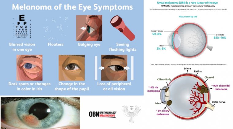
The retina receives blood supply from two vascular systems: the choroid, which primarily nourishes photoreceptor cells, and the central retinal artery, supplying the inner retinal layers. These systems differ significantly in blood flow rates and oxygenation levels, emphasizing the importance of precise measurement locations during retinal microcirculation assessments.
Evolution of Retinal Vessel Assessment
Since the invention of the ophthalmoscope in 1851 by Helmholtz, advancements in assessing retinal microcirculation have been substantial. Today, tools are available to measure vessel diameter, wall thickness, and blood flow velocity using techniques like laser Doppler flowmetry (LDF) and scanning laser Doppler flowmetry (SLDF).
LDF, utilizing a helium-neon laser, measures erythrocyte flow and tissue perfusion units. SLDF extends this capability to include arteriole morphology assessments, while bidirectional laser Doppler flowmetry (BLDV) offers comprehensive erythrocyte velocity evaluations.
Multiwavelength Laser Doppler Holography (MLDH)
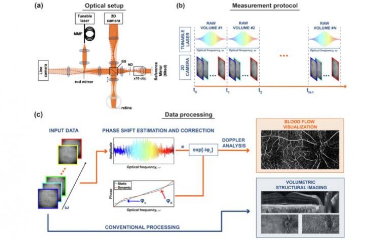
The introduction of MLDH enhances conventional STOC-T tomography by enabling the visualization and quantification of blood flow across retinal layers. This novel approach integrates laser flowmetry with holographic multiwavelength detection, delivering high-resolution, real-time assessments of blood flow dynamics.
Dr. Dawid Borycki explains, "Our method enables the acquisition of two-dimensional blood flow images from a stack of interferometric images with different wavelengths in approximately 8.5 milliseconds. This is comparable to the time needed for conventional optical OCT to register a pair of repeated cross-sectional scans."
Crucially, MLDH integration with STOC-T requires no modifications to existing protocols, leveraging the same dataset for enhanced diagnostic capabilities across retinal microcirculation assessments.
This breakthrough promises to advance clinical understanding of ocular and systemic health, providing clinicians with a comprehensive tool to monitor and diagnose circulatory disorders effectively.
Reference
Dawid Borycki et al, Multiwavelength laser doppler holography (MLDH) in spatiotemporal optical coherence tomography (STOC-T), Biocybernetics and Biomedical Engineering (2024). DOI: 10.1016/j.bbe.2024.03.002