(1).jpg)

New Digital Refocusing Method Could Simplify and Expand Access to Retinal Imaging
A collaborative research team from Johns Hopkins University, Boston University, and partners has developed a novel imaging system that enables digital refocusing during retinal exams, potentially reducing cost, complexity, and accessibility barriers associated with traditional fundus cameras.
Challenges in Conventional Fundus Imaging
Fundus imaging, the process of photographing the back of the eye, is a critical diagnostic tool in identifying major causes of vision loss such as diabetic retinopathy, glaucoma, and age-related macular degeneration (AMD). However, traditional fundus cameras require precise mechanical focusing and are typically expensive and difficult to operate, particularly in underserved clinics or remote healthcare settings.
A Computational Alternative to Mechanical Focus
In a study published in Biophotonics Discovery, researchers introduced a diffuser-based computational imaging system that removes the need for manual or mechanical focusing during eye exams. The system works by replacing a portion of a commercial fundus camera’s optics with a holographic diffuser and a high-sensitivity digital sensor.
This approach allows the camera to capture 3D light field information. The resulting data is then processed by computer algorithms to digitally refocus the retinal image after it has been captured, eliminating the need for lens adjustment during imaging.
First Human Demonstration and Imaging Performance
This study marks the first successful demonstration of this technology in living human eyes. After calibrating the system by measuring how the diffuser interacts with varying levels of refractive error, researchers were able to digitally correct the blur and reconstruct sharp images of the retina.
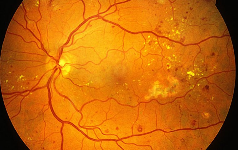
In clinical tests, the camera captured color images revealing key anatomical features such as the optic disc, blood vessels, and macula. Remarkably, the system could refocus images across more than 10 diopters of refractive error, without any moving optical components. The resolution remained stable at approximately 7 to 10 line pairs per millimeter, slightly lower than standard cameras but highly consistent, even in eyes with significant refractive error.
Implications for Accessibility and Integration
The findings demonstrate that diffuser-based computational refocusing is a viable method for use in real-world eye exams. Because it eliminates the need for mechanical focus mechanisms, this innovation could lead to the development of more affordable and accessible fundus cameras, especially in resource-limited environments.
Additionally, researchers suggest that this technique could be integrated with autorefractors, devices that measure eyeglass prescriptions, to create a combined diagnostic tool. Such a system would streamline comprehensive eye exams and broaden their availability in both primary care and global health settings.
Reference:
Corey Simmerer et al, In vivo fundus imaging and computational refocusing with a diffuser-based fundus camera, Biophotonics Discovery (2025). DOI: 10.1117/1.BIOS.2.4.042306