(1).jpg)
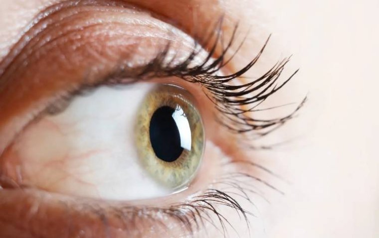
Landmark Genetic Study Reveals How the Fovea Develops to Enable Sharp Central Vision
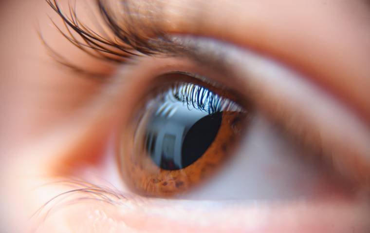
A study combining artificial intelligence and large-scale genetic analysis has provided the first comprehensive map of how the fovea, the small but vital part of the retina responsible for sharp central vision, develops in the human eye. Led by the University of Leicester and the Ulverscroft Eye Unit at University Hospitals of Leicester NHS Trust, in collaboration with multiple international institutions, the study marks a significant advance in understanding the genetic and developmental underpinnings of both rare and common vision disorders.
First Genome-Wide Study of Human Foveal Development
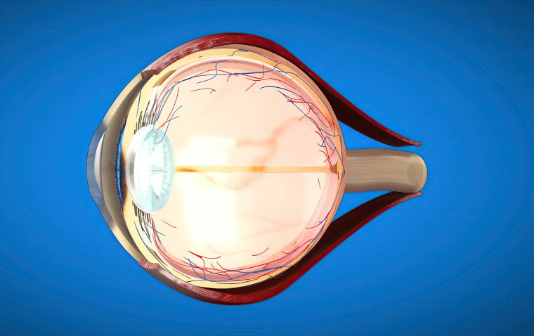
The research team conducted the first genome-wide association study (GWAS) of human foveal development, using advanced AI algorithms to analyze optical coherence tomography (OCT) scans from over 60,000 participants in the UK Biobank. The study was funded by the Wellcome Trust, the Ulverscroft Foundation, and the NIHR Leicester Biomedical Research Centre, among others.
Results were recently published in Investigative Ophthalmology & Visual Science and revealed over 120 genetic signals involved in foveal formation. Among them were 64 genes not previously associated with the fovea, uncovering novel biological pathways related to vitamin A metabolism, retinal cell differentiation, vascular development, and pigmentation.
Genetic Findings Expand Clinical Understanding
This new genetic map shows that both common and rare genetic variants influence whether the fovea forms correctly, shedding light on why developmental disorders like albinism, aniridia, and other genetic syndromes lead to foveal underdevelopment and long-term visual impairment.
The study also found that foveal abnormalities are not limited to ocular-specific conditions. They were identified in systemic disorders such as Stickler syndrome, Refsum disease, Leber congenital amaurosis with early-onset deafness, and microcephaly–chorioretinopathy syndromes, significantly broadening the relevance of these findings.
“The findings provide the first comprehensive genetic dissection of human foveal pit architecture, revealing entirely new biological mechanisms that shape foveal development and extending our understanding of childhood visual disorders,” said Dr. Mervyn Thomas, Clinical Associate Professor and Clinical Lead at the Ulverscroft Eye. “Our data highlights the role of retinoic acid (a vitamin A derivative) signaling in human foveal development—something previously unknown. These discoveries fundamentally change our understanding of how the back of the eye develops and open up new avenues for diagnosing and treating childhood vision loss.”
The study proposes a continuum model of eye development, suggesting that a spectrum of genetic variation, from subtle common variants to rare disease-causing mutations, can impact foveal structure and visual outcomes.
This research not only enhances understanding of the biological mechanisms behind high-acuity vision but also lays the foundation for future diagnostic tools, gene-targeted therapies, and precision medicine approaches for childhood and inherited eye diseases.
Reference:
Callum Hunt et al, Genome-Wide Insights Into the Genes and Pathways Shaping Human Foveal Development: Redefining the Genetic Landscape of Foveal Hypoplasia, Investigative Ophthalmology & Visual Science (2025). DOI: 10.1167/iovs.66.12.22