(1).jpg)
Alzheimer's First Signs May Appear in Your Eyes, New Study Finds
Researchers at Cedars-Sinai have conducted the most comprehensive analysis of retinal changes in Alzheimer’s disease patients and their correlation with brain and cognitive changes. The findings of this study, which have been published in the journal Acta Neuropathologica, are crucial for gaining a better understanding of the complex effects of Alzheimer's on the retina, particularly in the early stages of cognitive impairment. This understanding is essential in the development of more effective treatments to halt the progression of the disease.
Annually, over 3 million Americans receive a diagnosis of Alzheimer's disease, which gradually deteriorates their memory and cognitive ability. Currently, there is no definitive diagnostic test for Alzheimer's disease, and the latest treatments only delay the progression of the disease, rather than halting it entirely.
“Our study is the first to provide in-depth analyses of the protein profiles and the molecular, cellular, and structural effects of Alzheimer’s disease in the human retina and how they correspond with changes in the brain and cognitive function,” said Maya Koronyo-Hamaoui, PhD, professor of Neurosurgery, Neurology, and Biomedical Sciences at Cedars-Sinai and senior author of the study. “These findings may eventually lead to the development of imaging techniques that allow us to diagnose Alzheimer’s disease earlier and more accurately and monitor its progression noninvasively by looking through the eye.”
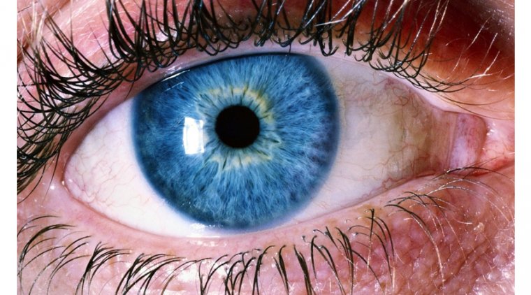
“The retina, a developmental extension of the brain, offers an unparalleled opportunity for affordable, noninvasive monitoring of the central nervous system,” said Yosef Koronyo, MSc, research associate in the Cedars-Sinai Department of Neurosurgery and first author of the study. “And with the help of our collaborators, we discovered the accumulation of highly toxic proteins in the retinas of patients with Alzheimer’s disease and mild cognitive impairment, causing severe degeneration of cells.”
The Most Extensive Analysis of the Retina: Key Findings
In the study, researchers analyzed retinal and brain tissue samples from 86 human donors, which were collected over a period of 14 years. This group of donors represents the largest sample of retinal tissue collected from patients with Alzheimer's disease and mild cognitive impairment. The samples were compared between donors with normal cognitive function, those with mild cognitive impairment at the earliest stages of Alzheimer's disease, and those with later-stage Alzheimer's disease dementia.
The researchers delved into the physical characteristics of the patients' retinas, measuring and mapping inflammation markers and functional cell loss. They also conducted an analysis of the proteins present in both retinal and brain tissues.
Here is what investigators found in the retinas of patients with mild cognitive impairment and Alzheimer’s disease:
● An overabundance of a protein called amyloid beta 42, which in the brains of Alzheimer’s disease patients clumps together to form plaques that disrupt brain function
● Accumulation of amyloid beta protein in ganglion cells, the cells that bridge visual input from the retina to the optic nerve
● Higher numbers of astrocytes and immune cells, called microglia, tightly surrounding amyloid beta plaques
● As many as 80% fewer microglial cells clearing amyloid beta proteins from the retina and brain
● Specific molecules and biological pathways responsible for inflammation, and cell and tissue death
“These changes in the retina correlated with changes in parts of the brain called the entorhinal and temporal cortices, a hub for memory, navigation, and the perception of time,” said Koronyo.
The changes observed in the retina were also linked to the pathological stage of Alzheimer's disease, commonly referred to as Braak stage, as well as the cognitive status of the patients. Interestingly, these changes were identified even in patients who were apparently cognitively normal or only very mildly impaired, suggesting that they could serve as an early predictor of subsequent cognitive decline.
“These findings give us a deeper understanding of the effects of Alzheimer’s disease on the retina,” said Keith L. Black, MD, chair of the Department of Neurosurgery and the Ruth and Lawrence Harvey Chair in Neuroscience at Cedars-Sinai and a co-author of the study. “Because these changes correspond with changes in the brain and can be detected in the earliest stages of impairment, they may lead us to new diagnostics for Alzheimer’s disease and a means to evaluate new forms of treatment.”
Reference
“Retinal pathological features and proteome signatures of Alzheimer’s disease” by Yosef Koronyo, Altan Rentsendorj, Nazanin Mirzaei, Giovanna C. Regis, Julia Sheyn, Haoshen Shi, Ernesto Barron, Galen Cook-Wiens, Anthony R. Rodriguez, Rodrigo Medeiros, Joao A. Paulo, Veer B. Gupta, Andrei A. Kramerov, Alexander V. Ljubimov, Jennifer E. Van Eyk, Stuart L. Graham, Vivek K. Gupta, John M. Ringman, David R. Hinton, Carol A. Miller, Keith L. Black, Antonino Cattaneo, Giovanni Meli, Mehdi Mirzaei, Dieu-Trang Fuchs and Maya Koronyo-Hamaoui, 11 February 2023, Acta Neuropathologica.
DOI: 10.1007/s00401-023-02548-2