Dry Eye Testing & New Diagnostic Tools
Dry eye disease (DED), also known as keratoconjunctivitis sicca, is one of the most common ophthalmic conditions, affecting hundreds of millions of people worldwide. Recent technological advances and research targeting DED have led to the emergence of new definitions and new approaches for DED diagnosis and management.
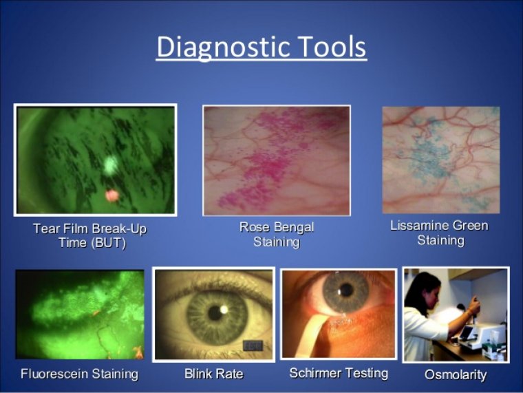
During the past several years, new diagnostic tools have revolutionized the face of dry eye disease (DED). Historically, DED diagnostic tools were limited to subjective grading of corneal staining, Schirmer tear tests, and counting tear breakup time (TBUT).
All provided us with some data on DED, yet DED is a dynamic condition in which signs do not always match symptoms and symptoms do not always match signs. Also, multiple factors can contribute to the disease severity.
Today, various available diagnostic tools help us sort out the key factors contributing to your patients’ DED, including point-of-care immunoassays that test for elevated levels of matrix metalloproteinase-9 (MMP-9) and tear osmolarity and imaging systems that evaluate the structural health of meibomian glands to help us to identify meibomian gland dysfunction (MGD).
All of these diagnostic tests evaluate different contributing factors for dry eye and assist physicians in their diagnosis and treatment of patients. Let’s review the diagnostic tests currently available.
Common Evaluation Tools
Historically, the most common evaluative tools included grading of corneal staining, Schirmer tear testing, and TBUT. Corneal staining is usually quantified using the standard grading system in ophthalmology (Trace, 1+, 2+, 3+, 4+).
Grading can vary based on the examiner and reflects general health of the ocular surface. Schirmer tear testing uses a paper strip placed in the lower eyelid after topical anesthetic is applied. After 5 minutes, the strip is read in millimeters. Less than 10 mm is considered dry, and greater than 10 mm is considered normal.
The SMTube (Quidel) is a similar test on the market. It is a strip meniscometry tube that replaces the Schirmer test. It takes 5 seconds to administer and does not require the use of anesthetic. It is also measured in millimeters: less than 5 mm is considered dry and greater than 5 mm is considered a normal result.
With TBUT, a fluorescein strip is moistened with saline and applied to the inferior cul-de-sac. Using the blue filter on the slit lamp, the patient is instructed to blink. The interval of time between the patient’s blink and the appearance of dry spots on the cornea represents the TBUT.
It is measured in seconds, and less than 10 seconds is considered a decreased TBUT. Although these tests can provide useful data points for the patient’s exam, they are not the most reliable or reproducible tests.
While they assist in evaluating severity of disease state, they don’t necessarily help our physicians sort out the specific causative factors of DED that assist in prescribing therapies and treatments.
Laboratory tests Recently on the market, two laboratory tests can help to guide therapeutic decision making in the office at the point of care: InflammaDry (Quidel) and TearLab Osmolarity System (TearLab Corporation).
They look at different biomarkers and therefore provide complementary information. InflammaDry evaluates for elevated MMP-9, which is an inflammatory biomarker present in approximately half of untreated symptomatic dry eye patients.
Elevated levels of MMP-9 cause epithelial cell disruption, which leads to corneal staining. It is also worth noting that elevated MMP-9 precedes corneal staining. The test is a lateral flow immunoassay and looks very similar to a pregnancy test. The result provided is either positive or negative.
By diagnosing inflammation on the ocular surface, we are identifying a previously sub-clinical finding that will guide therapeutic decision making in real time. The other laboratory test available is the TearLab Osmolarity System.
Tear osmolarity is best described as the salt content of the tears. When osmolarity is elevated, it represents decreased aqueous and contributes to epithelial cell death. This device utilizes disposable test chips on a handheld collection device and a docking station that reads the results.
Osmolarity is measured in milliosmoles/liter (mOsm/L) of solution. A normal reading falls below 300 mOsm/L. TearLab classifies mild dry eye as represented by a reading of 300-320 mOsm/L, moderate 320-340 mOsm/L, and severe as greater than 340 mOsm/L.
Also, an inter-eye difference of 8 mOsm/L or more is considered an abnormal result and reflects tear film instability.
Evaluating The Meibomian Glands
Additional diagnostic tests evaluate the health and functioning of the meibomian glands. The Meibomian Gland Evaluator (Johnson & Johnson Vision) is a handheld tool that assists in evaluating meibomian gland secretions.
This small device simulates the pressure of a blink, while the examiner looks for oil secretions from the meibomian gland orifices. Absence of secretion indicates that the meibomian glands are not functioning correctly.
Meibography is another useful tool. This test uses infrared imaging of the meibomian glands to evaluate the structure of the meibomian glands from the palpebral conjunctiva. Healthy meibomian glands appear tightly packed, straight, and numerous.
As MGD progresses, the patient experiences meibomian gland drop out, because the glands become truncated and less numerous. Once a gland has atrophied completely, its function is permanently lost.
Box Medical Solutions offers a portable, slit lamp mountain meibographer. The LipiScan with Dynamic Meibomian Imaging (DMI) (Johnson & Johnson Vision) provides high-definition imaging of meibomian gland structure.
The LipiView II Ocular Surface Interferometer with DMI (Johnson & Johnson Vision) and the Oculus Keratograph 5M (Oculus) are multi-modal devices that evaluate numerous findings related to DED and ocular surface health.
The LipiView II measures lipid layer thickness, captures blink dynamics, and images meibomian gland structure.
While the Oculus Keratograph 5M is an advanced corneal topographer, it also has the ability to perform meibography, evaluate the lipid layer, and measure TBUT as well as the tear meniscus height.
Enhancing patient care The advent of new technologies for dry eye patients has revolutionized the way practices evaluate their patients. These tests provide complementary information to help physicians sort out the specific causative factors for DED.
With this information, eye-care providers can aid their patients with the most appropriate therapies, at an earlier interval, and ultimately enhance the quality of patient care in the dry eye practice.
(1).jpg)
