(1).jpg)
Novel Imaging Method Holds Potential for Uveitis Monitoring and Prediction
Researchers at the Eye Clinic of the University Hospital Bonn (UKB) and the University of Bonn have found a promising imaging monitoring method utilizing optical coherence tomography angiography (OCTA). The technique assesses blood flow in retinal vessels, revealing crucial insights into the severity of inflammation and providing clues about the future course of uveitis.
Published in Scientific Reports, the study explores how this innovative approach could revolutionize uveitis monitoring, identifying patients at risk of disease worsening and potentially preventing complications leading to blindness.
Dr. Maximilian Wintergerst from the UKB Eye Clinic emphasizes the significance of early detection of uveitis, a rare disease with serious consequences, contributing to 5% to 10% of global blindness. The research focuses on intermediate uveitis, where inflammation primarily affects the vitreous body and, as indicated by preliminary work, can extend to the retinal vessels.
Recognizing the need for objective parameters to assess disease activity, the researchers investigated high-resolution imaging-based methods to enhance the understanding of uveitis. Dr. Wintergerst highlights the potential for "objective markers of inflammatory activity" to improve clinical monitoring and act as quantitative endpoints for future clinical trials.
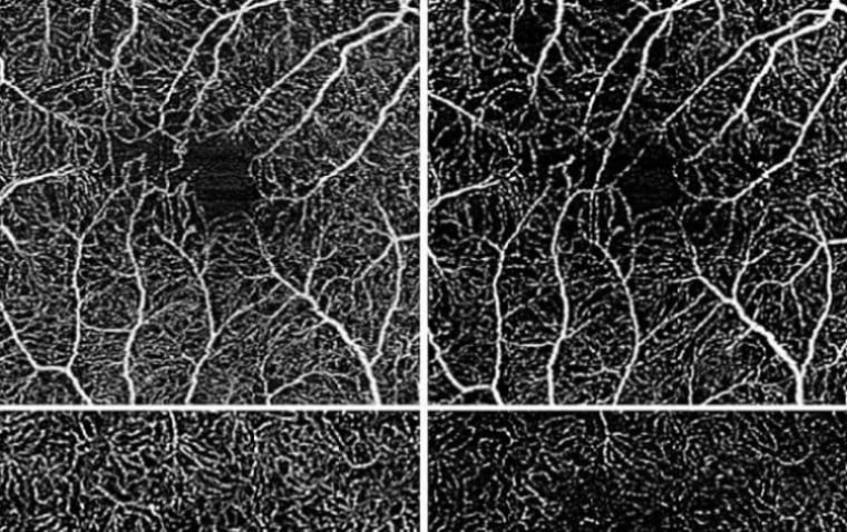
Optical coherence tomography angiography, a non-invasive and non-contact imaging method, emerged as a key tool. Dr. Wintergerst explains that the technique utilizes harmless laser light to scan the retina and choroid, generating tomographic images. By analyzing blood flow density, the researchers observed significant differences among eyes with stable disease, increased disease activity, and decreased disease activity.
The study, involving 52 participants, revealed a correlation between disease activity and blood flow density. Increased disease activity corresponded with decreased blood flow density, while decreased disease activity saw an increase in blood flow density.
To assess predictive power, the researchers employed a statistical model, analyzing more than 300 eye examinations. The results indicated that reduced blood flow density was significantly linked to future deterioration in central visual acuity.
Prof. Dr. Dr. Robert Finger, co-author of the study and Director of the Eye Clinic at the University Medicine Mannheim (UMM), envisions the data's potential to identify high-risk patients early on, enabling closer monitoring. He suggests using this parameter as an endpoint in future clinical trials to bolster evidence for treating this rare disease.
Prof. Dr. Frank Holz, Director of the UKB Eye Clinic, underscores the study's importance in establishing objective parameters for uveitis activity, paving the way for enhanced monitoring in the future. This novel imaging method holds promise for early intervention and improved outcomes for patients with uveitis.
Reference
Vessel density on optical coherence tomography angiography is prognostic for future disease course in intermediate uveitis, Scientific Reports (2024). DOI: 10.1038/s41598-023-49926-0