(1).jpg)

Dual Mechanisms Behind Rapid Eye Dominance Plasticity Discovered in Adult Brain
A new study published in Communications Biology has uncovered two distinct yet interconnected mechanisms responsible for ocular dominance plasticity in the adult brain. The research, led by a collaborative team from the Institute of Biophysics of the Chinese Academy of Sciences and Wenzhou Medical University, offers significant insight into how the adult visual system adapts to changes in binocular input, potentially informing future therapies for visual disorders like amblyopia.
High-Resolution fMRI Sheds Light on Ocular Plasticity
Using ultra-high field 7T functional MRI, the researchers investigated how short-term monocular deprivation affects ocular dominance in adults. Participants underwent just three hours of monocular contrast deprivation, during which one eye was deprived of high-contrast input.
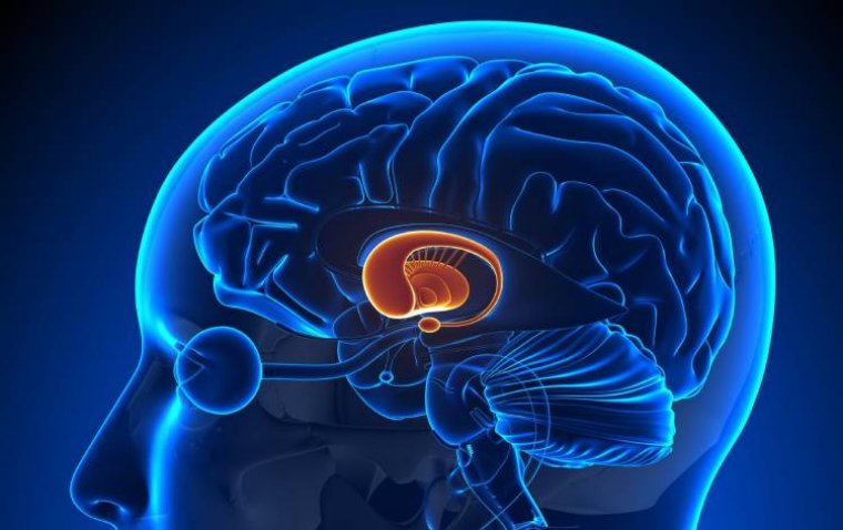
The imaging revealed a notable enhancement in both contrast sensitivity and ocular dominance in the deprived eye. This was associated with increased neural activity in the parvocellular layers of the lateral geniculate nucleus (LGN) and the ventrolateral subdivision of the pulvinar, key subcortical structures involved in visual processing.
The Non-Deprived Eye Shows Enhanced 3D Perception
Conversely, the non-deprived eye demonstrated significant improvements in 3D shape perception, along with heightened activation in stereopsis-related areas of the visual cortex. These changes highlight how both eyes undergo distinct yet coordinated adaptive responses when normal binocular input is disrupted.
A Reciprocal Pattern of Plasticity
One of the most striking findings of the study was the reciprocal relationship between plasticity in the pulvinar and the visual cortex. As activity increased in subcortical regions related to the deprived eye, there was a corresponding enhancement in cortical activity associated with the non-deprived eye. This pattern suggests two opposing yet complementary mechanisms that work together to maintain binocular balance.
Implications for Treating Visual Disorders
The discovery that the adult brain retains flexible and dual-site ocular dominance plasticity could be a game-changer in the field of ophthalmology. These mechanisms allow for rapid and adaptive visual system responses to short-term imbalances in input, offering promising implications for treating conditions like amblyopia.
By identifying distinct neural responses at both cortical and subcortical levels, this study opens the door to more targeted interventions that harness the brain’s natural plasticity to restore or enhance visual function in adults.
Reference:
Yazhu Qian et al, Two opposing yet complementary ocular dominance plasticities: thalamus strengthens the weak channel while higher cortex listens to the strong signal, Communications Biology (2025). DOI: 10.1038/s42003-025-08914-y