Yamane Technique - Intrascleral IOL Fixation
The ideal situation in any cataract surgery is with implantation of an intraocular lens (IOL). However, it does not always happen. This could be the result of an intraoperative complication or due to a congenital anomaly of the lens or following trauma.
Various techniques have been described in the literature from anterior chamber IOLs (AC-IOL) to retro-fixated iris-claw IOLs (RIC-IOL) to scleral-fixated IOLs (SF-IOL). SF-IOLs are the most popular approach, with its long-term stability superior to RIC-IOLs.
This could be due to a lack of long-term follow-up of RIC-IOLs. SF-IOLs, being stationed posterior to the iris plane, also avoid corneal decompensation seen in AC-IOLs and cause minimal or no iris trauma.
They also can be done where iris is damaged (due to the primary surgery), where RIC-IOL cannot be done.
Various surgical approaches have also been described for SF-IOL, of which the most popular techniques include sutured SF-IOL, Prolene Haptic (3-piece IOL) exteriorization with tucking of haptics in the scleral pocket, glued IOL, Yamane technique, and the X-NIT technique.
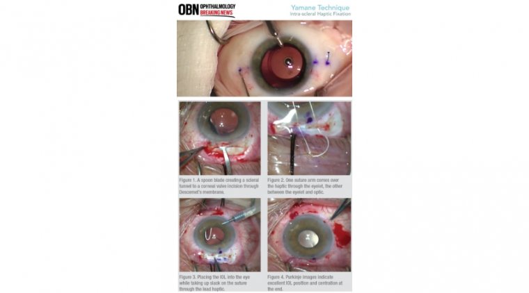
Yamane's technique of flanged intrascleral intraocular lens (IOL) fixation has become an accepted method of scleral fixating an IOL in eyes with suboptimal capsular support. The technique involves exteriorizing the haptics with 30-gauge needles through two angled incisions parallel to the limbus.
The haptics of the IOL are then cauterized to make a flange which is pushed back and fixed into the scleral tunnels. A disposable thermal cautery is recommended for creating the flange.
The Yamane technique, first described by Shin Yamane and colleagues, is of use for intrascleral IOL fixation when a patient is aphakic without capsular support. The technique offers an elegant and effective way to fixate the IOL in complex cases.
Thorough preparation, including practising in two wet labs before the fi rst case, is a must, and the learning curve with the Yamane technique can be steep.
However, it is also short. And whilst mastering a method usually requires a large number of cases, we have achieved great outcomes quickly by going back and reviewing videos and analysing my early experiences.
Patient cases – We were referred a patient with a posterior capsule tear during cataract surgery whose IOL had slipped nasally half inside and half outside the bag. The patient could no longer see well because the optic edge was now in the centre of the visual axis.
IOL exchange was scheduled. The dislocated IOL was carefully removed from the capsular bag and then cut into three pieces using 19g Packer/ Chang IOL cutters (MicroSurgical Technology [MST]) for atraumatic removal from the anterior chamber.
Following IOL removal, I inserted a CT Lucia lens (Zeiss) and both haptics into the anterior chamber. I then sutured the main incision (which was larger than usual because of the IOL removal) with the aim of avoiding excess fluid leakage and maintaining the pressure required for needle placement.
We used a 23g Ahmed micro-grasper (MST) through the paracentesis for entry to grasp the haptics. Placing the second haptic inside the anterior chamber made grasping and threading the haptic into the lumen of the needle much more challenging because the haptic slid into the angle.
With hindsight, we should have left the trailing haptic outside of the eye; we will do so in the future. In the next case, the patient’s lens had fallen into the posterior chamber during cataract surgery and the capsule was completely lost.
During the pars plana vitrectomy and lensectomy, they endured a sector iridectomy. The patient presented to us with aphakia, no capsule, some loss of iris tissue and cystoid macular oedema. We again elected to proceed with scleral fi xation utilising the Yamane technique.
This time, we used an AC maintainer included in the scleral IOL fi xation solutions pack (SFP1001, MST) to keep the eye firm. The needle placements went well, and there was no fl uid egress even with the trailing haptic exposed through the primary incision.
The procedure went smoothly and as planned. The trailing haptic can also secure the IOL from falling back into the vitreous cavity, especially in this case because there was no capsular support and iris tissue was missing.
We proceeded with the iris repair, which required suturing of the sphincter. We used 23g Ahmed micro-tying forceps (MST) to hold each edge of the tear as I passed the needle (10-0 prolene suture on CTC-6 needle) through to complete the repair.
At the end of the case, we saw that the IOL was not perfectly centred even though we had been careful with the scleral measurements, so we pulled the haptic to one side to centre the optic. We then trimmed the excess haptic material and mushroomed and reburied the tip.
The IOL was perfectly centred and there was no tilt at all. Learning points Measurements and markings Sometimes it can be difficult to mark accurately because the limbus is not always clearly defined.
One can miss by half a millimetre due to difficulty in identifying a clear entry point. In this case, even though one marks at 2.5 mm, this may not actually be 2.5 mm from the limbus. We believe this is what happened during the second case described above when we realised that the IOL was not well centred.
One needle entered further from the limbus than the other, which affected the IOL positioning. Of note, each needle pass is performed by a different hand and at a different angle—it is difficult to be perfectly symmetrical but awareness of the issue certainly helps.
Visibility - To know exactly where the positioning and entry points are (2.5 mm posterior to the limbus and 2 mm across), we like to open the conjunctiva and measure directly on the sclera to see as much as we can.
Surgeons who have experience with this technique can go through the conjunctiva, but we feel it is less stressful when you have better visibility to watch the needle pass under the sclera before making the turn.
The sclera can also be held to prevent the eye from rotating, which helps with the needle tunnelling. If going directly through conjunctiva, the scleral tunnel is not visible.
Finally, when turning the needle to enter the eye, go deep and not shallow: turn and go straight down and then come back up to visualise the bevel of the needle.
Right versus left hand - Keep in mind that the needles will be passed by both hands and that each hand will act differently: one comes forward while the other goes back.
There are going to be differences between your hand positions, which is not a big problem, but one need to be mindful of this and make adjustments to get the same result from both hands.
This is something that can be practiced with simulation model eyes. Fixation of the globe is very important; otherwise the eye rotates, so the hand positions should be thought out ahead of time.
Threading the haptic One might think that the haptic should thread directly into the needle, but in my experience, it is the opposite: threading the haptic is much easier if the needle is aimed toward the haptic while holding the haptic still.
We use the 30g thin-wall needles with specialist holders from the scleral IOL fixation solutions pack (MST), which allow for manoeuvrability and control during this delicate step.
To manipulate the haptic, our preferred micro-instrument is the 23g Ahmed micro-grasper (MST), which is a versatile micro-instrument for the anterior chamber.
Equipment and IOLs My final recommendation would be to choose your cases wisely and have retina support available. In addition, always prepare your equipment in advance. We opt for the CT Lucia IOL (Zeiss) with PVDF haptics, which I find has the right proportion of flexibility and resistance to breaking.
We also use the scleral IOL fixation solutions pack (MST), which includes the essential elements to perform the technique, including three 30g thin-wall needles with silicone hubs, two specialist needle holders, an AC maintainer, a marker and a calliper.
The Yamane technique can be easy to learn if one is prepared and well equipped. Even if things do not go perfectly the first couple of times, practice will help you learn from your mistakes and it will get easier.