
Laser eye surgery has become a popular solution for correcting vision problems such as nearsightedne...
read more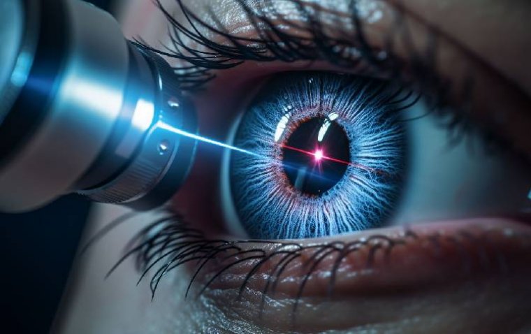
Ocular inflammation is a critical eye condition that can lead to discomfort, impaired vision, and, i...
read more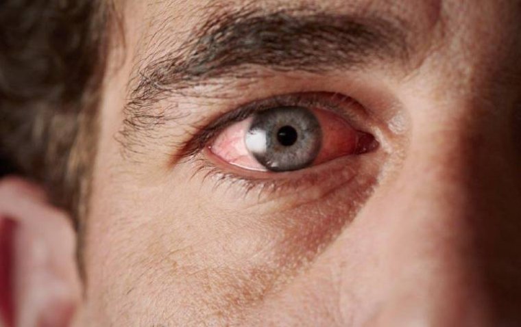
The ability of 3D bioprinting to fabricate complex tissue constructs holds the promise of overcoming...
read more
Smoking is widely recognized as a major health hazard, contributing to a range of systemic diseases,...
read more
A Freedom of Information (FOI) request has brought to light a concerning issue: thousands of blind a...
read more
In this article, we spotlight 10 organizations that stand out as heroes in the field of eye health, ...
read more
Eye color, a key trait that contributes to the uniqueness of an individual’s appearance, is determin...
read more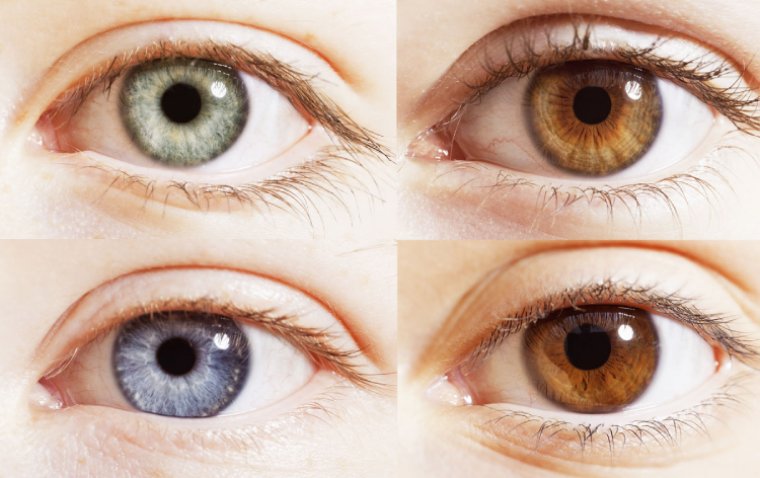
Experiencing sudden or temporary loss of vision in one eye can be a distressing event, signaling an ...
read more
In the modern workplace, maintaining eye health has become increasingly crucial, especially as many ...
read more
During the period of Ramadan, questions often arise about how certain health practices, such as the ...
read more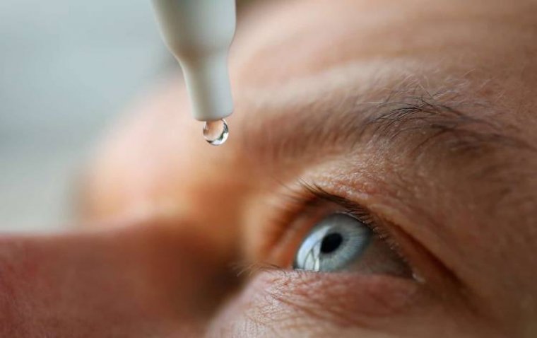
 More
More